. 2014 Jan 30;9(1):e86790.
doi: 10.1371/journal.pone.0086790. eCollection 2014.

Identification of STAT5A and STAT5B target genes in human T cells

Affiliations

Identification of STAT5A and STAT5B target genes in human T cells

Takahiro Kanai et al. PLoS One. .

Abstract

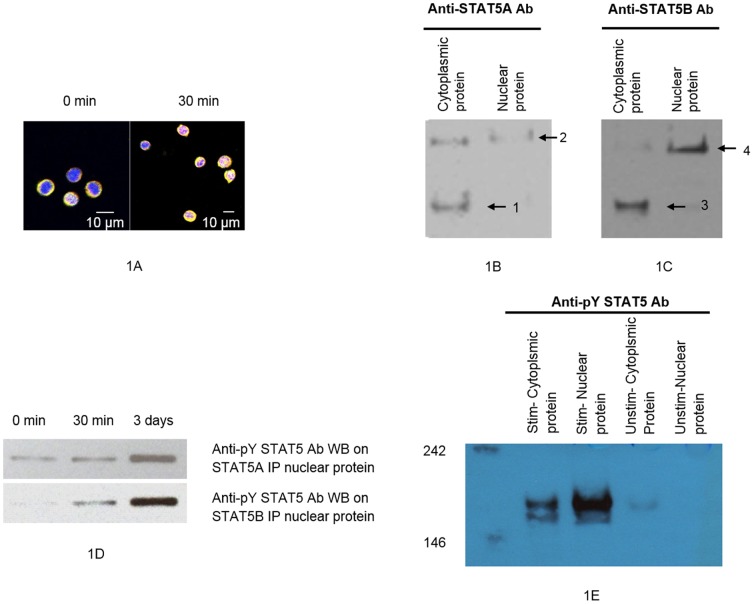
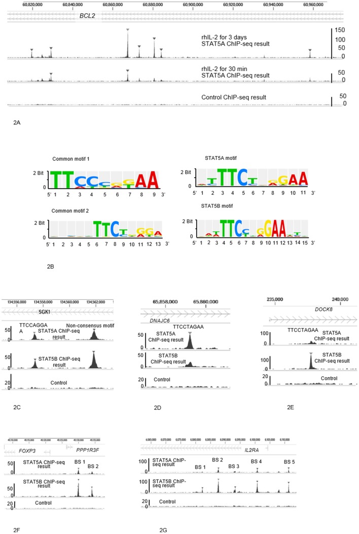
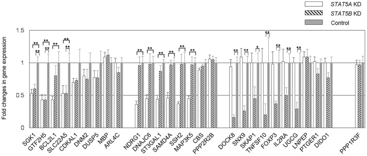
Signal transducer and activator of transcription (STAT) comprises a family of universal transcription factors that help cells sense and respond to environmental signals. STAT5 refers to two highly related proteins, STAT5A and STAT5B, with critical function: their complete deficiency is lethal in mice; in humans, STAT5B deficiency alone leads to endocrine and immunological problems, while STAT5A deficiency has not been reported. STAT5A and STAT5B show peptide sequence similarities greater than 90%, but subtle structural differences suggest possible non-redundant roles in gene regulation. However, these roles remain unclear in humans. We applied chromatin immunoprecipitation followed by DNA sequencing using human CD4(+) T cells to detect candidate genes regulated by STAT5A and/or STAT5B, and quantitative-PCR in STAT5A or STAT5B knock-down (KD) human CD4(+) T cells to validate the findings. Our data show STAT5A and STAT5B play redundant roles in cell proliferation and apoptosis via SGK1 interaction. Interestingly, we found a novel, unique role for STAT5A in binding to genes involved in neural development and function (NDRG1, DNAJC6, and SSH2), while STAT5B appears to play a distinct role in T cell development and function via DOCK8, SNX9, FOXP3 and IL2RA binding. Our results also suggest that one or more co-activators for STAT5A and/or STAT5B may play important roles in establishing different binding abilities and gene regulation behaviors. The new identification of these genes regulated by STAT5A and/or STAT5B has major implications for understanding the pathophysiology of cancer progression, neural disorders, and immune abnormalities.


Conflict of interest statement

Competing Interests: The authors declare no conflict of interest.

Figures

Figure 1
Figure 1. Localization of STAT5A and STAT5B, and monomers and dimers of STAT5A and STAT5B.
A demonstrates translocation of STAT5A and STAT5B into cell nuclei after 30-2 (40× confocal). Yellow, STAT5A; purple, STAT5B; blue, nucleus. B and C. Detection of STAT5A and STAT5B proteins in cytoplasmic or nuclear proteins fractionated from CD4+ T cells after PHA-P stimulation for 3 days followed by incubation with rhIL-2 for 30 min. B. STAT5A monomer (91 kDa, arrow 1) and STAT5A dimer (arrow 2) in native cytoplasmic or nuclear proteins, detected with anti-STAT5A Ab. C. STAT5B monomer (90 kDa, arrow 3) and STAT5B dimer (arrow 4) in native cytoplasmic or nuclear proteins, detected using anti-STAT5B Ab. D. Detection of phosphorylated STAT5 proteins in STAT5A- or STAT5B- immunoprecipitated nuclear proteins fractionated from CD4+ T cells after PHA-P stimulation for 3 days followed by incubation with rh-IL-2 for 0 min, 30 min or 3 days. E. Detection of phosphorylated STAT5 proteins in cytoplasmic or nuclear proteins fractionated from CD4+ T cells after PHA-P stimulation for 3 days followed by incubation with rhIL-2 for 3 days, and control (unstimulated condition).
Figure 2
Figure 2. Binding ability, motif sequences and binding sites.
A shows binding ability of STAT5A after 3 days of exposure to rhIL-2 versus binding ability of STAT5A after 30 min of exposure to rhIL-2. It shows results of STAT5A ChIP-seq on chromosome 18 performed in CD4+ T cells incubated with rhIL-2 for 3 days (top); in CD4+ cells incubated with rhIL-2 for 30 min (middle); compared with control ChIP-seq in CD4+ T cells (bottom). B shows consensus motif sequences for STAT5A and/or STAT5B. C shows binding sites for SGK1, detected by both STAT5A and STAT5B ChIP-seq. D and E show the detection of the sequence “TTCCTAGAA” by STAT5A ChIP-seq in DNAJC6, and by STAT5B ChIP-seq in DOCK8. F shows that FOXP3 and PPP1R3F are located within 10,000 bp of the two binding sites. G shows the five binding sites for IL2RA.
Figure 3
Figure 3. Validation of candidate genes from ChIP-seq via QT-PCR in STAT5A and STAT5B knock-down human CD4+ T cells.
The expression levels were compared STAT5A KD or STAT5B KD CD4+ T cells with control CD4+ T cells. Genes regulated similarly by both STAT5A and STAT5B are SGK1, GTF2H5 and SLC22A5. Genes regulated specifically by STAT5A are NDRG1, DNAJC6, ST3GAL1, SAMD4A, SSH2, MAP3K5 and BCL2L1. Genes regulated specifically by STAT5B are DOCK8, SNX9, SKAP1, TNFSF10, FOXP3, IL2RA and UGCG. Data is presented as mean ± SEM. *, P<0.01; **, P<0.001. There were no statistical differences if not annotated. Abbreviations: SGK1, serum/glucocorticoid regulated kinase 1; GTF2H5, general transcription factor IIH, polypeptide 5; BCL2L1, BCL2-like 1; SLC22A5, solute carrier family 22 (organic cation/carnitine transporter), member 5; CDKAL1, CDK5 regulatory subunit associated protein 1-like 1; DNM2, dynamin 2; DUSP5, dual specificity phosphatase 5; MBP, myelin basic protein; ARL4C, ADP-ribosylation factor-like 4C; NDRG1, N-myc downstream regulated 1; DNAJC6, DnaJ (Hsp40) homolog, subfamily C, member 6; ST3GAL1, ST3 beta-galactoside alpha-2,3-sialyltransferase 1; SAMD4A, sterile alpha motif domain containing 4A; SSH2, slingshot protein phosphatase 2; MAP3K5, mitogen-activated protein kinase kinase kinase 5; CBS, cystathionine-beta-synthase; PPP2R2B, protein phosphatase 2, regulatory subunit B, beta; DOCK8, dedicator of cytokinesis 8; SNX9, sorting nexin 9; SKAP1, src kinase associated phosphoprotein 1; PTGER1, prostaglandin E receptor 1 (subtype EP1), 42 kDa; DIDO1, death inducer-obliterator 1; TNFSF10, tumor necrosis factor (ligand) superfamily, member 10; FOXP3, forkhead box P3; IL2RA, interleukin 2 receptor, alpha; UGCG, UDP-glucose ceramide glucosyltransferase; LNPEP, leucyl/cystinyl aminopeptidase; PPP1R3F, protein phosphatase 1, regulatory subunit 3F.

Similar articles

Cited by

References

    1. Lin JX, Leonard WJ (2000) The role of Stat5a and Stat5b in signaling by IL-2 family cytokines. Oncogene 19: 2566–2576. - PubMed
    1. Rochman Y, Spolski R, Leonard WJ (2009) New insights into the regulation of T cells by gamma(c) family cytokines. Nat Rev Immunol 9: 480–490. - PMC - PubMed
    1. Leonard WJ, O'Shea JJ (1998) Jaks and STATs: biological implications. Annu Rev Immunol 16: 293–322. - PubMed
    1. Soldaini E, John S, Moro S, Bollenbacher J, Schindler U, et al. (2000) DNA binding site selection of dimeric and tetrameric Stat5 proteins reveals a large repertoire of divergent tetrameric Stat5a binding sites. Mol Cell Biol 20: 389–401. - PMC - PubMed
    1. Lin JX, Mietz J, Modi WS, John S, Leonard WJ (1996) Cloning of human Stat5B. Reconstitution of interleukin-2-induced Stat5A and Stat5B DNA binding activity in COS-7 cells. J Biol Chem 271: 10738–10744. - PubMed

Publication types