. 2014 Jan 30;9(1):e86843.
doi: 10.1371/journal.pone.0086843. eCollection 2014.

Preclinical evaluation of engineered oncolytic herpes simplex virus for the treatment of pediatric solid tumors

Affiliations

Preclinical evaluation of engineered oncolytic herpes simplex virus for the treatment of pediatric solid tumors

Michael L Megison et al. PLoS One. .

Abstract

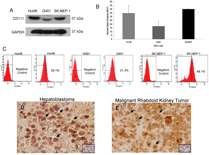
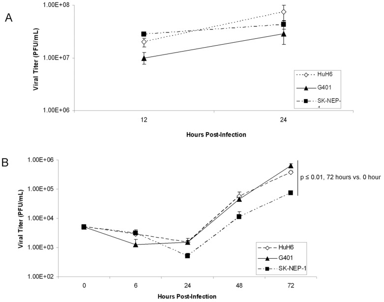
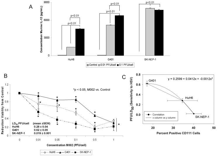
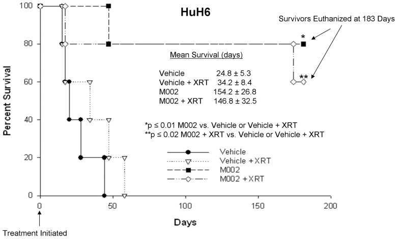
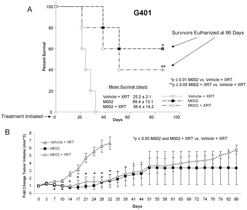
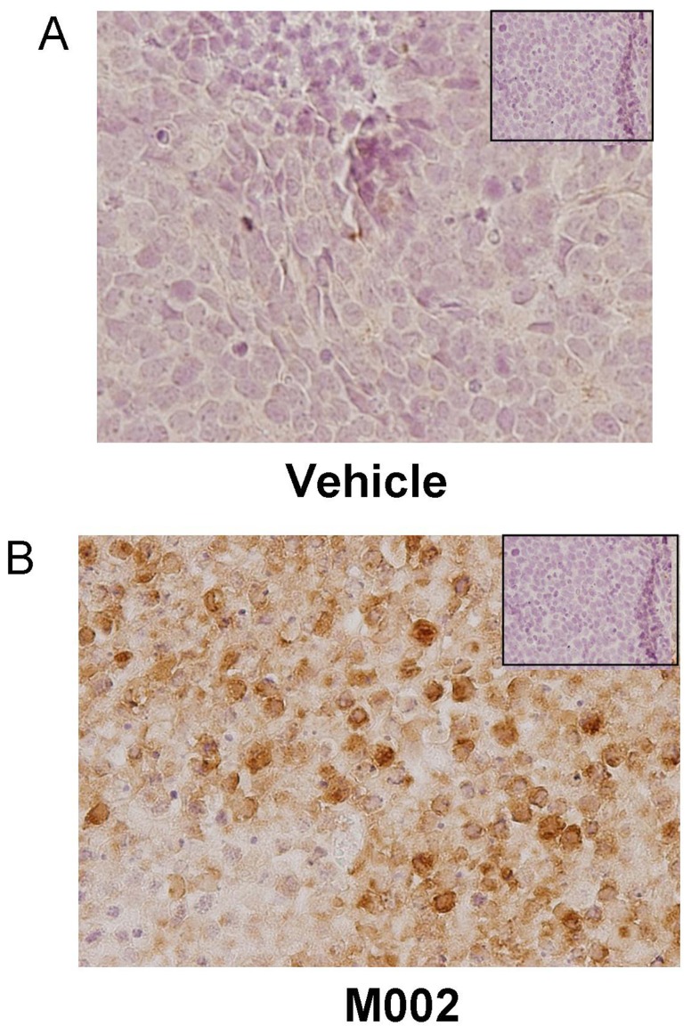
Recently, investigators showed that mice with syngeneic murine gliomas that were treated with a neuroattenuated oncolytic herpes simplex virus-1 (oHSV), M002, had a significant increase in survival. M002 has deletions in both copies of the γ134.5 gene, enabling replication in tumor cells but precluding infection of normal cells. Previous studies have shown antitumor effects of other oHSV against a number of adult tumors including hepatocellular carcinoma and renal cell carcinoma. The purpose of the current study was to investigate the oncolytic potential of M002 against difficult to treat pediatric liver and kidney tumors. We showed that the oHSV, M002, infected, replicated, and decreased cell survival in hepatoblastoma, malignant rhabdoid kidney tumor, and renal sarcoma cell lines. In addition, we showed that in murine xenografts, treatment with M002 significantly increased survival and decreased tumor growth. Finally, these studies showed that the primary entry protein for oHSV, CD111 (nectin-1) was present in human hepatoblastoma and malignant rhabdoid kidney tumor specimens. We concluded that M002 effectively targeted these rare aggressive tumor types and that M002 may have potential for use in children with unresponsive or relapsed pediatric solid tumors.


Conflict of interest statement

Competing Interests: The authors have declared that no competing interests exist.

Figures

Figure 1
Figure 1. CD111 expression in hepatoblastoma and rare pediatric renal tumor cell lines and specimens.
A Immunoblotting with CD111 specific antibody demonstrated that CD111 was present in whole cell lysates of HuH6, G401, and SK-NEP-1 cell lines. Detection of GAPDH was used to confirm equal protein loading. B HuH6, G401, and SK-NEP-1 cells were stained with fluorescence antibody for CD111. CD111 staining in these cell lines was detected in amounts that were consistent with those seen by immunoblotting (A). Stained cells were counted with flow cytometry and representative scatterplots presented for both negative controls and stained cells (C). D Immunohistochemistry staining with CD111 antibody was performed on 9 formalin-fixed, paraffin-embedded human hepatoblastoma and 12 malignant rhabdoid kidney tumor specimens. Representative photomicrographs presented at 40×, and negative controls were included with each (small box inserts). There was no CD111 staining detected in the negative controls. D Cellular staining for CD111 (black arrows) was present in over half of the hepatoblastoma specimens examined and was not limited to background stromal staining. E Staining for cellular CD111 (black arrows) was detected in the majority of MRKT specimens.
Figure 2
Figure 2. M002 infection.
A Single-step in vitro replication of M002. Monolayers of HuH6, G401 or SK-NEP-1 cells were infected with M002 at a MOI of 10 PFU/cell. Replicate cultures were harvested at 12 and 24 hours post-infection and virus titers determined on Vero cell monolayers. Mean virion yields were determined in four replicates at each time point and standard error of the mean was determined. B Multi-step replication of M002. Monolayers of HuH6, G401 or SK-NEP-1 cells were infected with M002 at a MOI of 0.1 PFU/cell, and at 6, 24, 48, 72 hours post-infection, supernates were collected and virus titers determined on Vero cell monolayers. Mean virion yields were determined in four replicates at each time point and standard error of the mean was determined. M002 replicated nearly 2 logs higher than control at 72 hours post-infection.
Figure 3
Figure 3. M002 infection and cell survival.
A To further verify infectivity, murine IL-12 production was determined in HuH6, G401 and SK-NEP-1 cell lines following treatment with M002 oHSV. Cell lines were infected with M002 at 0, 0.01 or 0.1 PFU/cell. After 48 hours, the supernates were collected and concentrations of murine IL-12 determined by ELISA. Data reported as mean ± standard error of the mean. There was a significant increase in murine IL-12 production in all cell lines even at the lower MOI of 0.01 PFU/cell. B HuH6, G401 and SK-NEP-1 cell lines were treated with M002 at increasing MOI. After 72 hr of treatment, cell viability was measured with alamarBlue™ assays. Data reported as mean ± standard error of the mean. There was a significant decrease in viability in all cell lines following M002 treatment. The LD50 was calculated for each cell line for M002 (PFU/cell). C A comparison was made between percent cells staining for CD111 and the sensitivity of the cell lines to M002. There was a non-linear, inverse correlation between the two variables. The straight lines represent the slope between two points and the curved line the correlation calculated with the three points, represented by the polynomial equation. The bars represent the SEM for the data points in both the x and y axis.
Figure 4
Figure 4. M002 treatment of hepatoblastoma xenografts.
HuH6 human hepatoblastoma tumor cells (2.5×10⁶ cells) in Matrigel™ were injected into the right flank of female athymic nude mice. Once tumors reached 250 mm3, animals received an intra-tumoral injection of vehicle [PBS +10% glycerol, 50 µL (n = 10)] or M002 virus [1×10⁷ PFU/50 µL (n = 10)]. Following injection, half of the animals in each group were treated with 3 Gy irradiation to the tumor. Kaplan-Meier graph for animal survival. Animals treated with M002 alone or with M002+ XRT had a significant increase in survival over those treated with vehicle alone or with vehicle+XRT. There was no significant survival advantage seen with the addition of XRT to either the vehicle treated animals or the M002 treated animals.
Figure 5
Figure 5
A The HuH6 tumor xenografts (as described in Figure 4) were weighed at euthanasia. The mean tumor weights from animals treated with M002 alone or M002+ XRT were significantly smaller than weights of the tumors from animals treated with vehicle or vehicle+XRT. Bars represent mean. Tumor weights were not altered by the addition of XRT to either the vehicle or M002 treatments. B HuH6 tumor xenograft volumes were measured biweekly with calipers [(width)2×length]/2. Data reported as mean fold change in tumor volume ± standard error. In the HuH6 xenografts, there was a significant decrease in tumor growth that began at day 7 following treatment that continued to the end of the study. The addition of low dose XRT to either the vehicle or M002 treatments had no significant effect upon tumor growth.
Figure 6
Figure 6. M002 treatment of malignant rhabdoid kidney tumor xenografts.
G401 human MRKT cells (2.5×10⁶ cells) in Matrigel™ were injected into the right flank of female athymic nude mice. Once tumors reached 250 mm3, animals received an intra-tumoral injection of vehicle [PBS +10% glycerol, 50 µL (n = 5)] or M002 virus [1×10⁷ PFU/50 µL (n = 10)]. Following injection, the vehicle treated and half of the M002 treated animals were given 3Gy irradiation to the tumor. A Kaplan-Meier graph for animal survival. Animals treated with M002 alone or with M002+ XRT had a significant increase in survival over those treated with vehicle+XRT. There was no significant survival advantage seen with the addition of XRT to the M002 treatment. B Tumor volumes were measured twice weekly [(width)2×length]/2. Data reported as mean fold change in tumor volume ± standard error. In the M002 treated G401 xenografts, there was a significant decrease in tumor growth that began at day 7 following treatment that continued to the end of the study. The addition of low dose XRT to M002 treatments had no significant effect upon tumor growth.
Figure 7
Figure 7. M002 treatment of renal Ewing sarcoma xenografts.
A SK-NEP-1 cells, (1.5×10⁶ cells) were injected into the subcapsular space of the left kidney in 6 week old female athymic nude mice. After three weeks, the renal tumors were directly injected with either control vehicle (PBS +10% glycerol/50 µL, n = 6) or M002 oncolytic herpes simplex virus (1×10⁷ PFU/50 µL, n = 7). Two weeks following treatment, the animals were euthanized and the kidney tumors were harvested, measured with a caliper and weighed. B Tumor volumes in the animals treated with M002 were half those of animals treated with vehicle only (bar = mean). C Tumor weights following M002 treatment were also nearly half of those of vehicle treated tumors (bar = mean). D Formalin-fixed, paraffin embedded samples of vehicle treated SK-NEP-1 tumor specimens were stained for hematoxylin and eosin. Examination of these kidney tumor specimens revealed native tumor architecture consisting of sheets of small round blue cells. Representative photomicrographs at 10× with enlarged area of detail at 40×. E Formalin-fixed, paraffin embedded samples of M002 treated SK-NEP-1 tumor specimens were stained for hematoxylin and eosin. These specimens showed tumor necrosis (open arrow) and hemorrhage (pink area), and inflammatory cell infiltrates (closed arrow). Representative photomicrographs at 10× with enlarged area of detail at 40×.
Figure 8
Figure 8. Immunohistochemical staining for HSV in SK-NEP-1 tumor xenografts.
Formalin-fixed, paraffin embedded samples of SK-NEP-1 tumor xenografts (those presented in the data in Figure 5) were stained for HSV using immunohistochemistry, and representative photomicrographs presented. A No HSV was detected by immunohistochemical staining in SK-NEP-1 tumors treated with vehicle alone. Negative controls (rabbit IgG) were included with each run (small box insert). B HSV immunohistochemical staining revealed HSV present in the M002 treated SK-NEP-1 xenografts (brown stain). There was no viral staining seen in the negative controls (rabbit IgG) (small box insert).

Similar articles

Cited by

References

    1. Stocker JT (1994) Hepatoblastoma. Semin Diagn Pathol 11: 136–143. - PubMed
    1. Giacomantonio M, Ein SH, Mancer K, Stephens CA (1984) Thirty years of experience with pediatric primary malignant liver tumors. J Pediatr Surg 19: 523–526. - PubMed
    1. Allan BJ, Parikh PP, Diaz S, Perez EA, Neville HL, et al... (2013) Predictors of survival and incidence of hepatoblastoma in the paediatric population. HPB (Oxford) doi: 10.1111/hpb.12112. [Epub ahead of print]. - PMC - PubMed
    1. Amar AM, Tomlinson G, Green DM, Breslow NE, de Alarcon PA (2001) Clinical presentation of rhabdoid tumors of the kidney. J Pediatr Hematol Oncol 23: 105–108. - PubMed
    1. Zhuge Y, Cheung MC, Yang R, Perez EA, Koniaris LG, et al. (2010) Pediatric non-Wilms renal tumors: subtypes, survival, and prognostic indicators. J Surg Res 163: 257–263. - PubMed

Publication types

MeSH terms