. 2014 Jan 30;9(1):e87145.
doi: 10.1371/journal.pone.0087145. eCollection 2014.

Peste des petits ruminants virus tissue tropism and pathogenesis in sheep and goats following experimental infection

Affiliations

Peste des petits ruminants virus tissue tropism and pathogenesis in sheep and goats following experimental infection

Thang Truong et al. PLoS One. .

Abstract

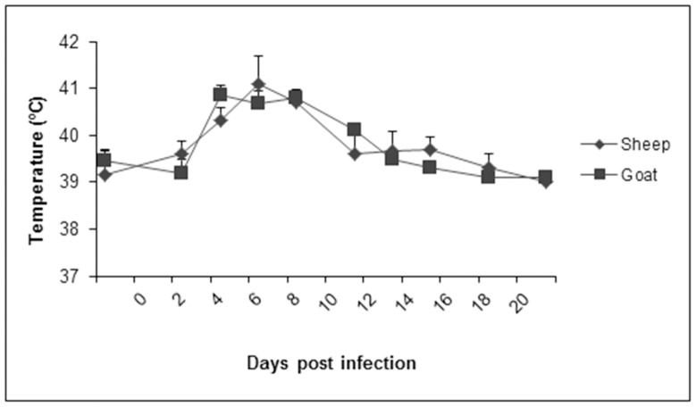
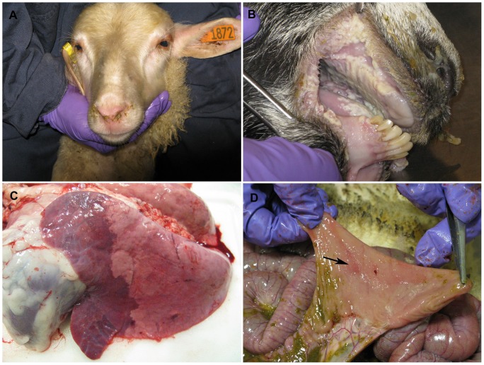
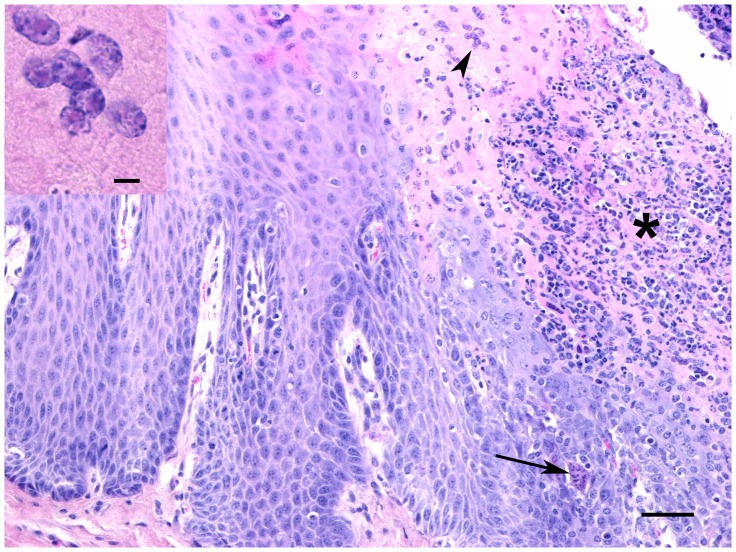
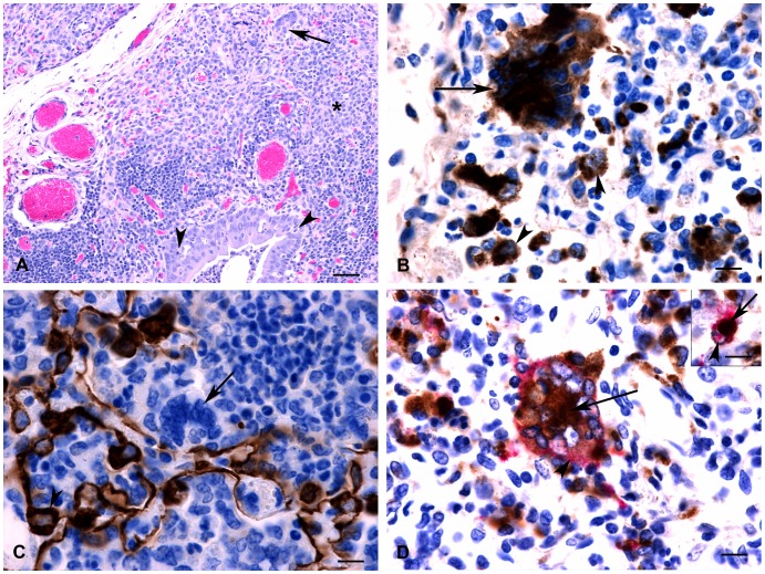
Peste des petits ruminants (PPR) is a viral disease which primarily affects small ruminants, causing significant economic losses for the livestock industry in developing countries. It is endemic in Saharan and sub-Saharan Africa, the Middle East and the Indian sub-continent. The primary hosts for peste des petits ruminants virus (PPRV) are goats and sheep; however recent models studying the pathology, disease progression and viremia of PPRV have focused primarily on goat models. This study evaluates the tissue tropism and pathogenesis of PPR following experimental infection of sheep and goats using a quantitative time-course study. Upon infection with a virulent strain of PPRV, both sheep and goats developed clinical signs and lesions typical of PPR, although sheep displayed milder clinical disease compared to goats. Tissue tropism of PPRV was evaluated by real-time RT-PCR and immunohistochemistry. Lymph nodes, lymphoid tissue and digestive tract organs were the predominant sites of virus replication. The results presented in this study provide models for the comparative evaluation of PPRV pathogenesis and tissue tropism in both sheep and goats. These models are suitable for the establishment of experimental parameters necessary for the evaluation of vaccines, as well as further studies into PPRV-host interactions.


Conflict of interest statement

Competing Interests: The authors have declared that no competing interests exist.

Figures

Figure 1
Figure 1. Rectal temperatures of sheep and goats following PPRV infection.
Rectal temperatures of sheep and goats were measured 2 days prior to experimental infection with PPRV (Malig strain), and following infection at regular intervals until 21 dpi. Results presented are the mean temperatures with standard deviations from animals at each time point.
Figure 2
Figure 2. Clinical signs and gross pathology in sheep and goats following infection with PPRV (Malig strain).
(A) Nasal discharges were observed in sheep at 6 dpi. (B) At 8 dpi, goats developed significant erosions of oral mucosa, as well as (C) bronchointerstitial pneumonia. (D) Depressed and reddened Peyer’s patches from infected sheep at 11 dpi.
Figure 3
Figure 3. Cross-section through a tonsil from a goat at 8 dpi.
There is necrosis of surface epithelium and extensive neutrophilic infiltrate (*) as well as occasional syncytial cells (arrow) and intranuclear inclusion bodies in upper epithelial layers (arrowhead, see inset). HE stain, bar = 50 µm. Inset: Eosinophilic intranuclear inclusion bodies. Bar = 5 µm.
Figure 4
Figure 4. Histology and immunohistochemistry of sheep and goat tissue at varying time points following infection with PPRV (Malig strain).
(A) Section of a lymph node; goat, 6 dpi. Multinucleated syncytial cells (arrows) and degenerating or apoptotic lymphocytes (arrowheads) were observed at 6 and 8 dpi. Inset: Higher magnification showing detail of apoptotic lymphocytes. HE stain, bar = 20 µm (B) Lymph node; goat, 6 dpi. Positive immunostaining using PPRV-specific antibodies in syncytial cells (arrows) and macrophages (arrowheads). Bar = 20 µm. (C) Section of omasum; sheep, 8 dpi. There is necrosis and loss of epithelium with edema, neutrophil infiltration (*) and syncytial cell formation (arrows). HE stain, bar = 50 µm. (D) Abomasum; goat, 8 dpi. Positive immunostaining for PPRV antigen could be detected within the gastric pits and glands as well as in the associated lymphoid tissue (*). Bar = 100 µm. (E) Ileum; sheep, 6 dpi. There is positive immunostaining for PPRV antigen within Peyer’s patches (arrow) as well as crypt epithelial cells (arrowhead). Bar = 50 µm. (F) Liver; sheep, 8 dpi. Focal area of hepatocyte loss with non-suppurative inflammation and degenerating syncytial cells (arrows). HE stain, bar = 50 µm.
Figure 5
Figure 5. Histopathology and immunohistochemistry of goat lung at 8 dpi.
(A) Hyperplasia of bronchiolar epithelium is evident with scattered epithelial degeneration (arrowheads) and abundant neutrophils within the lumen. Surrounding parenchyma is consolidated (*) with severe infiltration of mononuclear inflammatory cells. Note large syncytial cell (arrow). (B) Positive immunolabelling for CD68 was observed within multinucleated syncytial cells indicating they are of monocyte/macrophage origin (arrow). Note positive immunostaining of adjacent macrophages (arrowheads). (C) Positive immunolabeling for cytokeratin is observed within pneumocytes (arrowhead); however, syncytial cells are negative (arrow) indicating that they are not of epithelial origin. (D) Double immunolabelling detected the simultaneous expression of CD68 macrophage marker (brown stain, arrow) and PPRV antigen (pink stain, arrowhead) within multinucleated syncytial cells. Inset: Double immunolabelling detected expression of CD68 macrophage marker (brown stain, arrowhead) and PPRV antigen (pink stain, arrow) within the same cell indicating the presence of viral antigen within macrophages. Bar = 10 µm.
Figure 6
Figure 6. Quantification of PPR viral RNA in blood, nasal and oral swabs determined using real-time RT-PCR.
Both nasal and oral swabs were collected from sheep (A) and goats (B) at various time points until 21 dpi. Viral RNA quantification from whole blood (C) was also performed at identical time points. Note that in the case of sheep (C), viral RNA was not detectable at any time point before or after experimental infection with PPRV. Results presented are the mean value with standard deviation from animals at each time point. P<0.05 for sheep and goat nasal swabs at 6, 8 and 11 dpi and for sheep and goat oral swabs at 6 and 8 dpi compared to −2 dpi by t-test. P<0.05 for goats whole blood at 4, 6 and 8 dpi compared to −2 dpi by t-test.
Figure 7
Figure 7. Seroconversion following experimental PPRV infection.
PPRV-specific antibody titres in serum from sheep (A) and goats (B) measured using an indirect ELISA and virus neutralization test (VNT). Results presented are the mean values with standard deviations from animals at each time point. P<0.05 for ELISA from sheep and goats starting at 8 dpi compared to −2 dpi and P<0.05 for VNT from sheep and goats starting at 11 dpi compared to −2 dpi by t-test.
Figure 8
Figure 8. Quantification of interferon-gamma (IFN-γ) in serum samples collected from sheep and goats following PPRV infection.
Measurement of IFN-γ levels were performed using an IFN-γ ELISA and cross-reactive antibodies against bovine IFN-γ, and quantified using a standard provided by the manufacturer. Results presented are the mean values with standard deviations from animals at each time point. P<0.05 for goats at 8 dpi compared to −2 dpi by t-test.

References

    1. Gargadennec L, Lalanne A (1942) La peste des petits ruminants. Bulletin des Services Zoo Techniques et des Epizzoties de l’Afrique Occidentale Francaise 5: 16–21.
    1. Wang Z, Bao J, Wu X, Liu Y, Li L, et al. (2009) Peste des petits ruminants virus in Tibet, China. Emerg Infect Dis 15: 299–301. - PMC - PubMed
    1. Ayari-Fakhfakh E, Ghram A, Bouattour A, Larbi I, Gribaa-Dridi L, et al. (2011) First serological investigation of peste-des-petits-ruminants and rift valley fever in Tunisia. Vet J 187: 402–404 10.1016/j.tvjl.2010.01.007;10.1016/j.tvjl.2010.01.007 - DOI - PubMed
    1. Diallo A, Minet C, Le Goff C, Berhe G, Albina E, et al. (2007) The threat of peste des petits ruminants: Progress in vaccine development for disease control. Vaccine 25: 5591–5597 10.1016/j.vaccine.2007.02.013 - DOI - PubMed
    1. African Union Internation Bureau for Animal Resources. (2012) Impact of livestock diseases in Africa. : 1–2–7.

Publication types

MeSH terms

LinkOut - more resources