Enhanced HBsAg synthesis correlates with increased severity of fibrosis in chronic hepatitis B patients
- PMID: 24498079
- PMCID: PMC3909099
- DOI: 10.1371/journal.pone.0087344
Enhanced HBsAg synthesis correlates with increased severity of fibrosis in chronic hepatitis B patients
Abstract
Background and aims: Little is known about whether low serum HBsAg levels result from impaired HBsAg synthesis or a reduced number of hepatocytes caused by advanced liver fibrosis. Therefore, we investigated the capacity for HBsAg synthesis in a cross-sectional cohort of treatment-naïve chronic hepatitis B patients.
Methods: Chronic hepatitis B patients (n = 362) were enrolled; liver biopsies were performed and liver histology was scored, and serum HBsAg and HBV DNA levels were investigated. In the enrolled patients, 183 out of 362 have quantitative serum HBsAg levels. Tissue HBsAg was determined by immunohistochemistry.
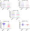
Results: A positive correlation between serum HBsAg and HBV DNA levels was revealed in HBeAg(+) patients (r = 0.2613, p = 0.0050). In HBeAg(+) patients, serum HBsAg and severity of fibrosis were inversely correlated (p = 0.0094), whereas tissue HBsAg levels correlated positively with the stage of fibrosis (p = 0.0280). After applying the mean aminopyrine breath test as a correction factor, adjusted serum HBsAg showed a strong positive correlation with fibrosis severity in HBeAg(+) patients (r = 0.5655, p<0.0001). The adjusted serum HBsAg values predicted 'moderate to severe' fibrosis with nearly perfect performance in both HBeAg(+) patients (area under the curve: 0.994, 95% CI: 0.983-1.000) and HBeAg(-) patients (area under the curve: 1.000, 95% CI: 1.000-1.000).
Conclusions: Although serum HBsAg levels were negatively correlated with fibrosis severity in HBeAg(+) patients, aminopyrine breath test-adjusted serum HBsAg and tissue HBsAg, two indices that are unaffected by the number of residual hepatocytes, were positively correlated with fibrosis severity. Furthermore, adjusted serum HBsAg has an accurate prediction capability.
Conflict of interest statement
Figures


References
-
- Chen CJ, Yang HI, Su J, Jen CL, You SL, et al. (2006) Risk of hepatocellular carcinoma across a biological gradient of serum hepatitis B virus DNA level. JAMA 295: 65–73. - PubMed
-
- Iloeje UH, Yang HI, Su J, Jen CL, You SL, et al. (2006) Predicting cirrhosis risk based on the level of circulating hepatitis B viral load. Gastroenterology 130: 678–686. - PubMed
-
- Chen YC, Sheen IS, Chu CM, Liaw YF (2002) Prognosis following spontaneous HBsAg seroclearance in chronic hepatitis B patients with or without concurrent infection. Gastroenterology 123: 1084–1089. - PubMed
-
- Yuen MF, Wong DK, Fung J, Ip P, But D, et al. (2008) HBsAg Seroclearance in chronic hepatitis B in Asian patients: replicative level and risk of hepatocellular carcinoma. Gastroenterology 135: 1192–1199. - PubMed
-
- European Association For The Study Of The Liver (2012) EASL clinical practice guidelines: Management of chronic hepatitis B virus infection. J Hepatol 57: 167–185. - PubMed
Publication types
MeSH terms
Substances
LinkOut - more resources
Full Text Sources
Other Literature Sources
Medical

