. 2014 Jan 30;9(1):e87350.
doi: 10.1371/journal.pone.0087350. eCollection 2014.

Crystal structure of an (R)-selective ω-transaminase from Aspergillus terreus

Affiliations

Crystal structure of an (R)-selective ω-transaminase from Aspergillus terreus

Andrzej Łyskowski et al. PLoS One. .

Abstract

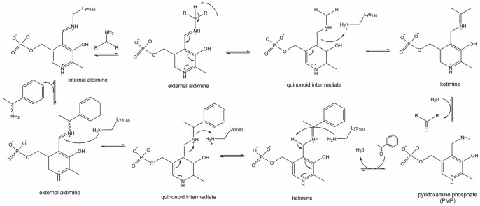
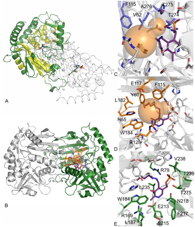
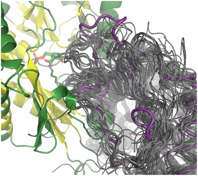
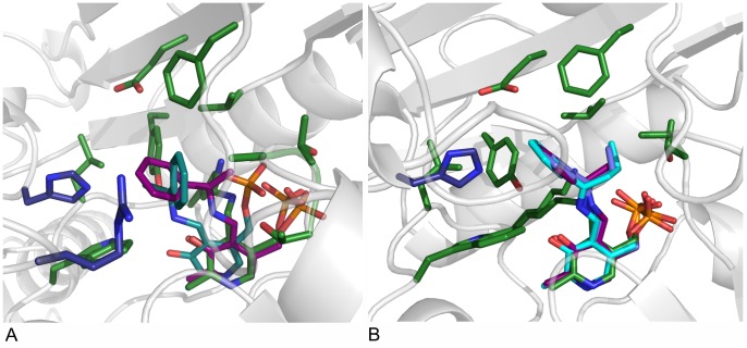
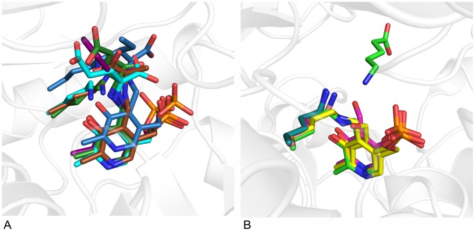
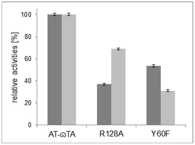
Chiral amines are important building blocks for the synthesis of pharmaceutical products, fine chemicals, and agrochemicals. ω-Transaminases are able to directly synthesize enantiopure chiral amines by catalysing the transfer of an amino group from a primary amino donor to a carbonyl acceptor with pyridoxal 5'-phosphate (PLP) as cofactor. In nature, (S)-selective amine transaminases are more abundant than the (R)-selective enzymes, and therefore more information concerning their structures is available. Here, we present the crystal structure of an (R)-ω-transaminase from Aspergillus terreus determined by X-ray crystallography at a resolution of 1.6 Å. The structure of the protein is a homodimer that displays the typical class IV fold of PLP-dependent aminotransferases. The PLP-cofactor observed in the structure is present in two states (i) covalently bound to the active site lysine (the internal aldimine form) and (ii) as substrate/product adduct (the external aldimine form) and free lysine. Docking studies revealed that (R)-transaminases follow a dual binding mode, in which the large binding pocket can harbour the bulky substituent of the amine or ketone substrate and the α-carboxylate of pyruvate or amino acids, and the small binding pocket accommodates the smaller substituent.


Conflict of interest statement

Competing Interests: Martin Schürmann is employed by a commercial company, DSM. This does not alter the authors’ adherence to all the PLOS ONE policies on sharing data and materials.

Figures

Figure 1
Figure 1. Detailed reaction mechanism of transaminases.
Figure 2
Figure 2. Crystal structure of AT-ωTA.
A: Overview of the AT-ωTA dimer (chain A in green, chain B in grey), conserved regions are indicated in yellow, B: overview of the AT-ωTA dimer with the binding pockets indicated as orange spheres, C: small binding pocket amino acids (blue), D: large binding pocket amino acids (orange), E: PLP binding amino acids (green). The figures were prepared using the program PyMOL.
Figure 3
Figure 3. Zoom into the loop Thr121-Val136 region of chain B in the structural alignment of the AT-ωTA structure (magenta) with other fold class IV transaminase structures (dark grey).
For PDB-IDs see Figure S2 in File S1. The figure was prepared using the program PyMOL.
Figure 4
Figure 4. Docking of various substrate intermediates into AT-ωTA.
A: Pro-(R)- and (S)-acetophenone pyridoxal phosphate intermediates docked into the active site of AT-ωTA. Green: amino acids of the active site (chain A) and PLP bound to K180, blue: amino acids of the active site (chain B), purple: pro-(R)-acetophenone pyridoxal phosphate intermediate, turquoise: pro-(S)-acetophenone pyridoxal phosphate intermediate. B: Acetophenone pyridoxal phosphate intermediate (purple), propiophenone pyridoxal phosphate intermediate (blue), butyrophenone pyridoxal phosphate intermediate (turquoise) docked into the active site of AT-ωTA compared to PLP bound to K180 in the structure of AT-ωTA (green). Green: amino acids of the active site (chain A), blue: amino acids of the active site (chain B). The figures were prepared using the program PyMOL.
Figure 5
Figure 5. Comparison of fold IV and fold I transaminases.
A: Position of lysine relative to PLP in fold IV transaminases: in AT-ωTA (green), BCAT from human (1KT8, blue) or E. coli (1IYE, turquoise) and D-ATA from Bacillus sp. YM-1 (3DAA, brown). Ligands: blue: L-Ile-aldimine bound in human BCAT, turquoise: L-Glu-aldimine bound in BCAT from E. coli, brown: D-Ala-aldimine bound in D-ATA, green: L-Glu-aldimine bound in AT-ωTA, purple: docked acetophenone-aldimine in AT-ωTA. B: Position of lysine relative to PLP in fold I (S)-ω-transaminases: in PD-ωTA from Paracoccus denitrificans (4GRX, light green), PA-ωTA from Pseudomonas aeruginosa (4B98, turquoise) and several (S)-TAs identified from the Pdb by Steffen-Munsberg: from Pseudomonas putida (3A8U, pink), from Mesorhizobium loti (3GJU, yellow) and from Silicobacter pomeroyi (3HMU, brownish), in PD-ωTA the substrate, 5-aminopentanoate, is depicted in light green. The figures were prepared using the program PyMOL.
Figure 6
Figure 6. Schematic drawing of the localisation of the large and small binding pocket in AT-ωTA relative to PO4 and O3’ of PLP and the binding of the substrates’ substituents.
Figure 7
Figure 7. Relative activities calculated from the increase of acetophenone at 300-ωTA and mutants thereof (0.1 mM PLP, 5 mM (R)-α-methylbenzylamine, 5 mM pyruvate or 5 mM butanal, in 50 mM KPi, pH 7.5, 0.25 mg/mL of total lysate protein) at 25°C.
The relative activities are referred to either the wild-type activity with pyruvate (0.8 U/mg lysate, dark grey bars) or butanal (0.1 U/mg lysate, light grey bars).

References

    1. Breuer M, Ditrich K, Habicher T, Hauer B, Kesseler M, et al. (2004) Industrial methods for the production of optically active intermediates. Angew Chem Int Ed 43: 788–824. - PubMed
    1. Höhne M, Bornscheuer U (2009) Biocatalytic routes to optically active amines. ChemCatChem 1: 42–51.
    1. Koszelewski D, Göritzer M, Clay D, Seisser B, Kroutil W (2010) Synthesis of optically active amines employing recombinant ω-transaminases in E. coli cells. ChemCatChem 2: 73–77.
    1. Carr R, Alexeeva M, Dawson M, Gotor-Fernandez V, Humphrey C, et al. (2005) Directed evolution of an amine oxidase for the preparative deracemisation of cyclic secondary amines. ChemBioChem 6: 637–639. - PubMed
    1. Ismail H, Lau R, van Rantwijk F, Sheldon R (2008) Fully enzymatic resolution of chiral amines: acylation and deacylation in the presence of Candida antarctica lipase B. Adv Synth Catal. 350: 1511–1516.

Publication types