. 2014 Jan 31;9(1):e87354.
doi: 10.1371/journal.pone.0087354. eCollection 2014.

β-hairpin-mediated formation of structurally distinct multimers of neurotoxic prion peptides

Affiliations

β-hairpin-mediated formation of structurally distinct multimers of neurotoxic prion peptides

Andrew C Gill. PLoS One. .

Abstract

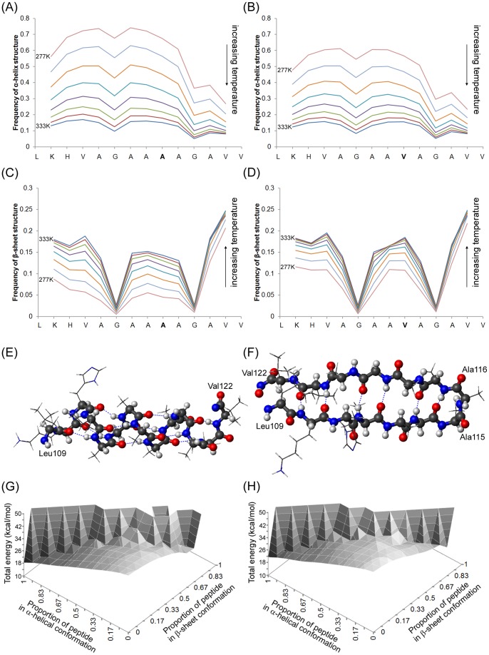
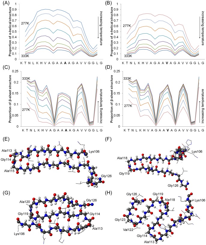
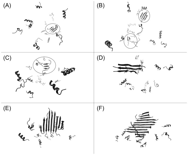
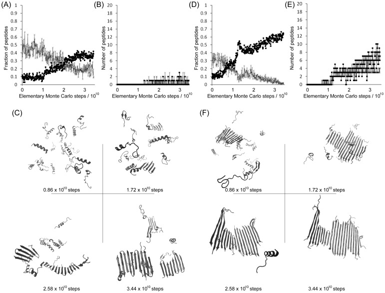
Protein misfolding disorders are associated with conformational changes in specific proteins, leading to the formation of potentially neurotoxic amyloid fibrils. During pathogenesis of prion disease, the prion protein misfolds into β-sheet rich, protease-resistant isoforms. A key, hydrophobic domain within the prion protein, comprising residues 109-122, recapitulates many properties of the full protein, such as helix-to-sheet structural transition, formation of fibrils and cytotoxicity of the misfolded isoform. Using all-atom, molecular simulations, it is demonstrated that the monomeric 109-122 peptide has a preference for α-helical conformations, but that this peptide can also form β-hairpin structures resulting from turns around specific glycine residues of the peptide. Altering a single amino acid within the 109-122 peptide (A117V, associated with familial prion disease) increases the prevalence of β-hairpin formation and these observations are replicated in a longer peptide, comprising residues 106-126. Multi-molecule simulations of aggregation yield different assemblies of peptide molecules composed of conformationally-distinct monomer units. Small molecular assemblies, consistent with oligomers, comprise peptide monomers in a β-hairpin-like conformation and in many simulations appear to exist only transiently. Conversely, larger assemblies are comprised of extended peptides in predominately antiparallel β-sheets and are stable relative to the length of the simulations. These larger assemblies are consistent with amyloid fibrils, show cross-β structure and can form through elongation of monomer units within pre-existing oligomers. In some simulations, assemblies containing both β-hairpin and linear peptides are evident. Thus, in this work oligomers are on pathway to fibril formation and a preference for β-hairpin structure should enhance oligomer formation whilst inhibiting maturation into fibrils. These simulations provide an important new atomic-level model for the formation of oligomers and fibrils of the prion protein and suggest that stabilization of β-hairpin structure may enhance cellular toxicity by altering the balance between oligomeric and fibrillar protein assemblies.


Conflict of interest statement

Competing Interests: The author has read the journal's policy and has the following conflicts: ACG is an academic editor for Plos One. This does not alter adherence to all the PLOS ONE policies on sharing data and materials.

Figures

Figure 1
Figure 1. Sequence of the murine prion protein.
The expressed protein has N- and C-terminal signal peptides (italics) that are removed during cellular processing. The N-terminal region is dynamically unstructured, whilst the C-terminal domain possesses globular structure, including two short sections of β-strand (bold text) with three α-helices (bold and underlined text). The hydrophobic peptide spanning residues 105–125 is highlighted in black with white text; the peptide spanning residues 108–121 is contained wholly within this peptide. Note, these peptides are homologous to human residues 106–126 and 109–122 and different numbering results from different lengths of the open reading frames. Throughout the rest of this paper the human numbering is used for consistency with other papers in the field.
Figure 2
Figure 2. Analysis of Monte Carlo simulations of 109–122 and 109–122 A117V peptide monomers.
(A) and (B) frequency of α-helical structure around each amino acid as a function of temperature for 109–122 and 109–122 A117V peptides respectively. Simulation temperatures are given in materials and methods and as the temperature is increased so the overall frequency of helical structure decreases. (C) and (D) frequency of β-sheet structure around each amino acid as a function of temperature for 109–122 and 109–122 A117V peptides respectively. As temperature increases the frequency of β-sheet also increases. (E) Schematic representation of the lowest energy conformation from the 3 replicate simulations of the wildtype 109–122 peptide. The backbone of the peptide is highlighted (carbon atoms grey, oxygen atoms red, nitrogen atoms blue) and the peptide is essentially 100% α-helical (F) schematic representation of the lowest energy conformer from the 3 replicate simulations of the wildtype 109–122 peptide in which β-sheet percentage is >50%. The peptide is in a β-hairpin conformation with a β-turn around residues Ala115 and Ala116. (G) and (H) total energy as a function of the proportion of helical structure and the proportion of β-sheet structure in 109–122 and 109–122 A117V mutant peptides respectively.
Figure 3
Figure 3. Analysis of Monte Carlo simulations of 106–126 and 106–126 A117V peptide monomers.
(A) and (B) frequency of α-helical structure around each amino acid as a function of temperature for 106–126 and 106–126 A117V peptides respectively. Simulation temperatures are given in materials and methods and as the temperature is increased so the overall frequency of helical structure decreases. (C) and (D) frequency of β-sheet structure around each amino acid as a function of temperature for 106–126 and 106–126 A117V peptides respectively. As temperature increases the frequency of β-sheet also increases. (E) Schematic representation of the 106–126 A117V peptide as a β-hairpin with a β-turn located around residues 113–115. The backbone of the peptide is highlighted (carbon atoms grey, oxygen atoms red, nitrogen atoms blue) (F) Schematic representation of the 106–126 A117V peptide as a β-hairpin with a β-turn around residues 118–119. (G) Schematic representation of the 106–126 A117V peptide as a double β-hairpin with β-turns around residues 113–114 and 119–120 (H) Schematic representation of the 106–126 A117V peptide as a triple β-hairpin conformation with β-turns around residues 113–114, 118–119 and 122–123. In all hairpin structures the β-turn incorporates one of the glycine residues of peptide 106–126.
Figure 4
Figure 4. In some multi-peptide simulations of the 109–122 peptide, large, multi-molecular assemblies are formed.
The data in this figure relates to replicate 1 at a simulation temperature of 293(A) fraction of peptide having α-helical structure (×) or β-sheet structure (▪) as a function of Monte Carlo steps. (B) The total energy of the system as a function of Monte Carlo steps. (C) the amount of peptides in parallel β-sheets (×) or in anti-parallel β-sheets (▪) as a function of Monte Carlo steps. (D) Ribbon representations of snapshots of the system at regular points throughout the simulation, as indicated.
Figure 5
Figure 5. Molecular assemblies that form during the pre- and mid-association phase of fibrillization of the 109–122 peptide.
(A–C) Ribbon representations of transiently stable multimers formed prior to nucleation of fibrillization. Dimers and trimers form that are often composed, partly, of peptides in β-hairpin conformation. (D) Ribbon representation of the trimer that represents the nucleus of fibril formation and is composed of peptides that have formed linear β-strands. (E–F) Ribbon representations of the growing fibril shortly after nucleation occurred. Peptides that have added to the ends of the growing fibril are often in β-hairpin conformation for a large number of Monte Carlo steps and inhibit addition of more peptides until the β-hairpin peptides open to form extended β-strands.
Figure 6
Figure 6. Structurally-different multimeric assemblies of the 106–126 peptide can form during simulations.
Panels (A–C) relate to replicate 1 at 293 K, whilst panels (D–F) relate to replicate 1 at 303 K. (A) and (D) show the fraction of peptide that possesses α-helical structure (×)or β-sheet structure (▪) as a function of Monte Carlo steps. (B) and (E) show the extent of oligomerization of peptide chains into parallel (×) or anti-parallel (▪) β-sheets as a function of Monte Carlo steps. (C) and (F) show ribbon representations of the state of the peptide ensemble at regular time points during each simulation. In panel (C), assemblies composed of peptides in β-hairpin conformation form roughly halfway through the simulation period. In panel (F), a nucleus of peptides in β-hairpin conformation form early during the simulation and, eventually, peptides being to form a fibril-like structure composed of β-strands.

References

    1. Chiti F, Dobson CM (2006) Protein misfolding, functional amyloid, and human disease. Annual Review of Biochemistry 75: 333–366. - PubMed
    1. Clark A, Dekoning EJP, Hattersley AT, Hansen BC, Yajnik CS, et al. (1995) Pancreatic Pathology in Non-Insulin-Dependent Diabetes (Niddm). Diabetes Research and Clinical Practice 28: S39–S47. - PubMed
    1. Goldschmidt L, Teng PK, Riek R, Eisenberg D (2010) Identifying the amylome, proteins capable of forming amyloid-like fibrils. Proceedings of the National Academy of Sciences of the United States of America 107: 3487–3492. - PMC - PubMed
    1. Laganowsky A, Liu C, Sawaya MR, Whitelegge JP, Park J, et al. (2012) Atomic View of a Toxic Amyloid Small Oligomer. Science 335: 1228–1231. - PMC - PubMed
    1. Yu LP, Edalji R, Harlan JE, Holzman TF, Lopez AP, et al. (2009) Structural Characterization of a Soluble Amyloid beta-Peptide Oligomer. Biochemistry 48: 1870–1877. - PubMed

Publication types

MeSH terms