. 2014 Feb 3;9(2):e87365.
doi: 10.1371/journal.pone.0087365. eCollection 2014.

TAF4 inactivation reveals the 3 dimensional growth promoting activities of collagen 6A3

Affiliations

TAF4 inactivation reveals the 3 dimensional growth promoting activities of collagen 6A3

Igor Martianov et al. PLoS One. .

Abstract

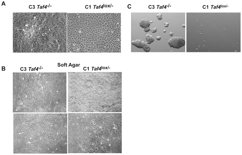
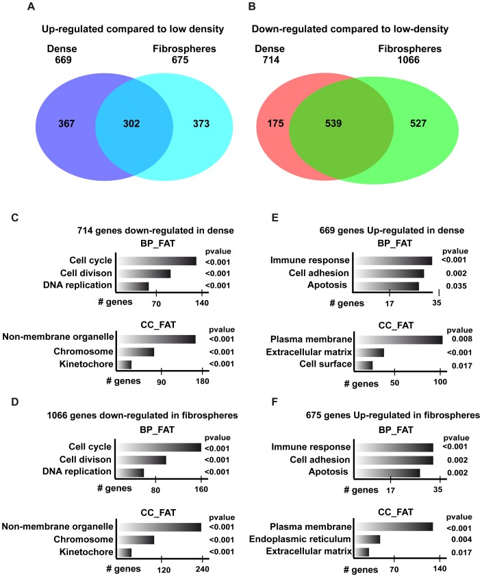
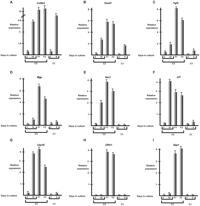
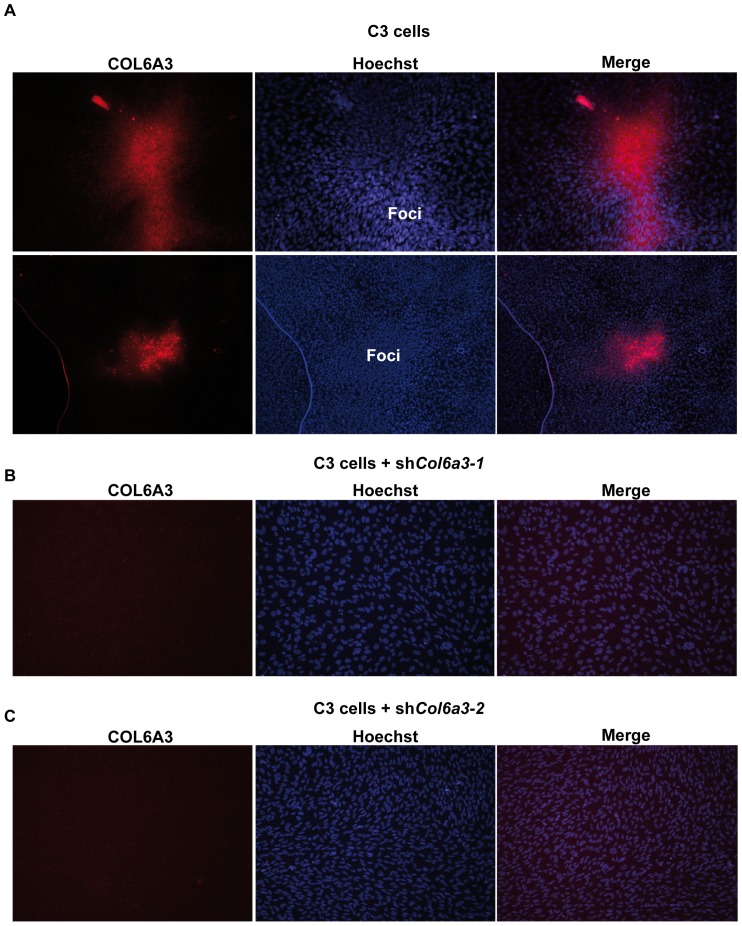
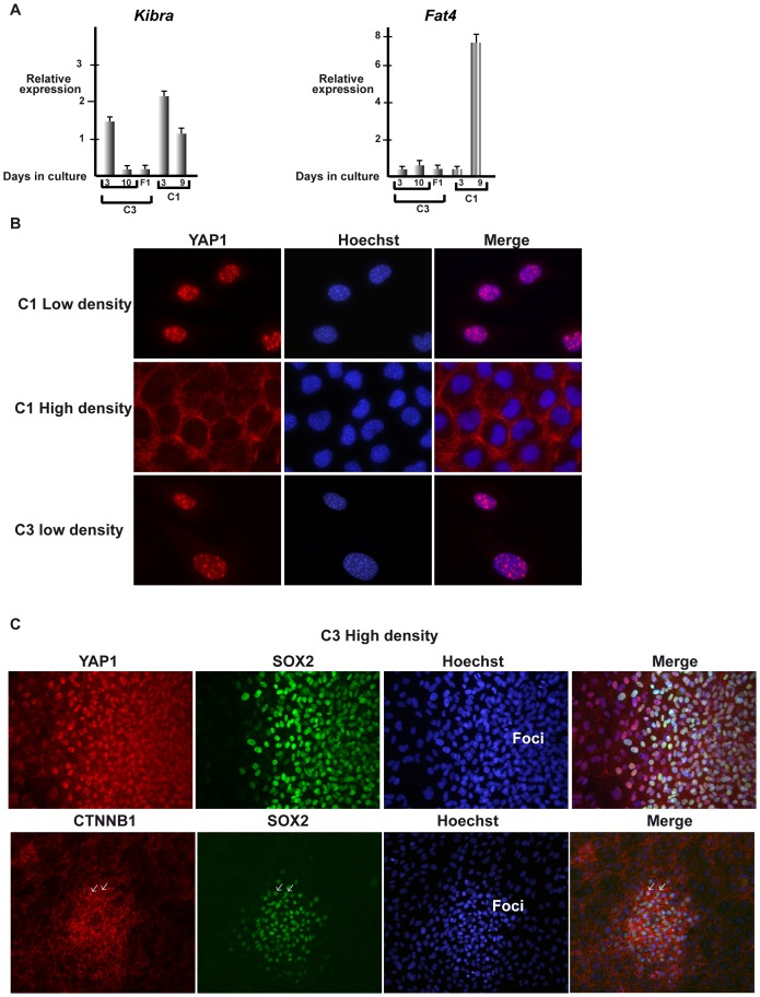
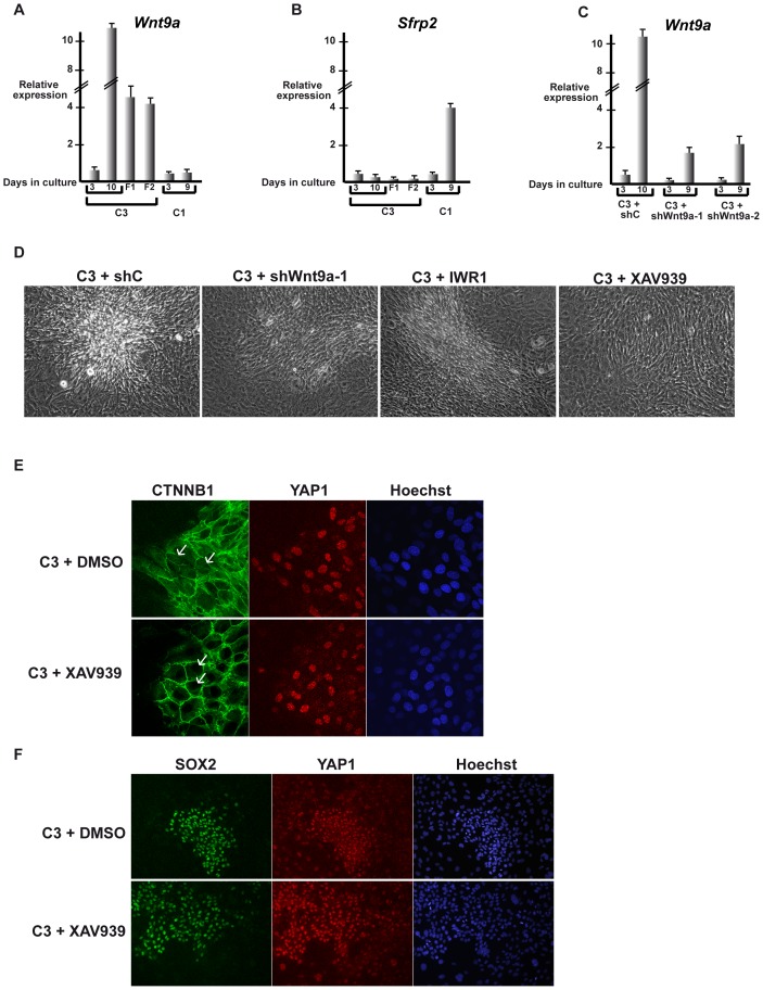
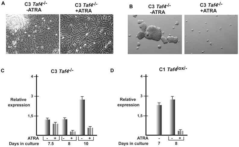
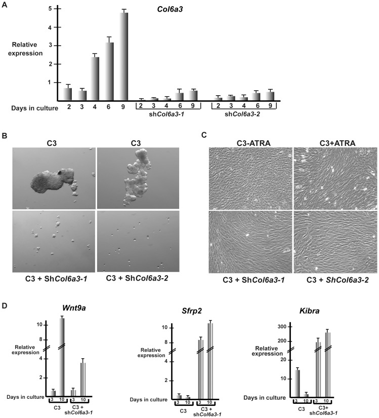
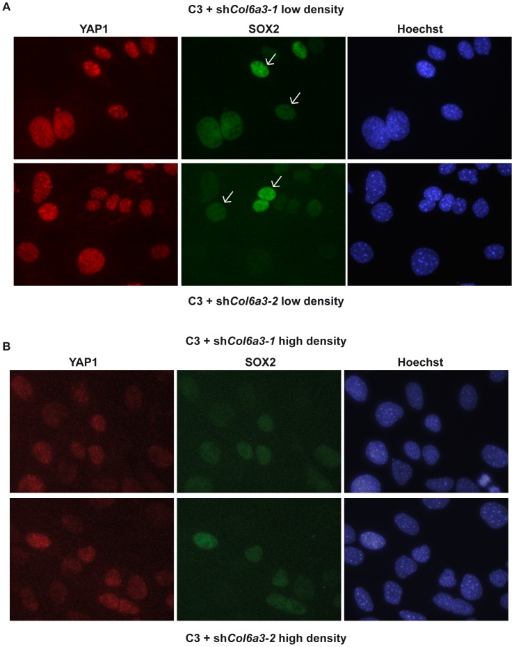
Collagen 6A3 (Col6a3), a component of extracellular matrix, is often up-regulated in tumours and is believed to play a pro-oncogenic role. However the mechanisms of its tumorigenic activity are poorly understood. We show here that Col6a3 is highly expressed in densely growing mouse embryonic fibroblasts (MEFs). In MEFs where the TAF4 subunit of general transcription factor IID (TFIID) has been inactivated, elevated Col6a3 expression prevents contact inhibition promoting their 3 dimensional growth as foci and fibrospheres. Analyses of gene expression in densely growing Taf4(-/-) MEFs revealed repression of the Hippo pathway and activation of Wnt signalling. The Hippo activator Kibra/Wwc1 is repressed under dense conditions in Taf4(-/-) MEFs, leading to nuclear accumulation of the proliferation factor YAP1 in the cells forming 3D foci. At the same time, Wnt9a is activated and the Sfrp2 antagonist of Wnt signalling is repressed. Surprisingly, treatment of Taf4(-/-) MEFs with all-trans retinoic acid (ATRA) restores contact inhibition suppressing 3D growth. ATRA represses Col6a3 expression independently of TAF4 expression and Col6a3 silencing is sufficient to restore contact inhibition in Taf4(-/-) MEFs and to suppress 3D growth by reactivating Kibra expression to induce Hippo signalling and by inducing Sfrp2 expression to antagonize Wnt signalling. All together, these results reveal a critical role for Col6a3 in regulating both Hippo and Wnt signalling to promote 3D growth, and show that the TFIID subunit TAF4 is essential to restrain the growth promoting properties of Col6a3. Our data provide new insight into the role of extra cellular matrix components in regulating cell growth.


Conflict of interest statement

Competing Interests: The authors have declared that no competing interests exist.

Figures

Figure 1
Figure 1. 3D growth of C3 cells.
A. Phase contrast microscopy (20× magnification) of C1 and C3 cells grown as dense cultures for 3 days. B. Growth of C1 and C3 cells after 10 days in soft agar. C. Phase contrast microscopy (12× magnification) of C1 cells or C3 cells in grown for 10 days under non-adherent conditions.
Figure 2
Figure 2. Changes in gene expression upon 3D growth of C3 cells.
A–B Venn diagrammes showing overlapping changes in gene expression upon growth as dense cultures or fibrospheres. C–F. Ontology analysis (http://david.abcc.ncifcrf.gov/) of the deregulated genes showing some relevant categories from the CC-FAT and BP FAT classifications. The number of genes and the associated P values for each category are indicated.
Figure 3
Figure 3. Comparative gene expression in C1 and C3 cells.
A–I. Results of RT-qPCR analysis of the indicated genes in C1 or C3 cells grown for 3 and 10 days in monolayer cluture or 10 days as fibrospheres (F1 and F2).
Figure 4
Figure 4. Elevated COL6A3 expression in MEFs growing as 3D foci.
A. Immunostaining of dense C3 MEFs for COL6A3. Two distinct fields are shown. COL6A3 is not homogeneously expressed, but strong expression is limited to the cells forming the 3D foci. B–C. Immunostaining of dense C3 cells expressing shRNAs directed against Col6a3 demonstrating the specificity of the staining.
Figure 5
Figure 5. Differential regulation of Hippo signalling in C1 and C3 MEFs.
A Results of RT-qPCR analysis of the indicated genes in C1 or C3 cells grown for 3 and 10 days in monolayer culture, or 10 days as fibrospheres (F1). B Immunostaining of C1 and C3 MEFs for YAP1 at low or high densities. C Immunostaining of C3 MEFs grown at high density for YAP1, SOX2 or β-catenin as indicated.
Figure 6
Figure 6. Wnt signalling is required for 3D growth.
A–C. Results of RT-qPCR analysis of the indicated genes in C1 or C3 cells grown for 3 and 9–10 days in monolayer cluture, 10 days as fibrospheres (F1 and F2) or in C3 cells expressing the indicated shRNAs. D Phase contrast microscopy (20× magnification) of C3 cells grown in presence or absence of the indicated Wnt pathway inhibitors or expressing control shRNA and shRNA directed againt Wnt9a. E. Confocal microscopy sections through foci grown in the presence or absence of XAV939 showing that the nuclear β-catenin localisation seen in the control C3 cells is lost in presence of XAV939. F. Immunostaining for YAP1 and SOX2 in foci in presence or absence of XAV939.
Figure 7
Figure 7. ATRA induced changes in properties of C3 MEFs.
A. Phase contrast microscopy (20× magnification) of C3 cells grown as dense cultures for 3 days in presence or absence of ATRA. B. Phase contrast microscopy (12× magnification) of C3 cells in grown for 10 days as fibrospheres in presence or absence of ATRA. CD Effect of ATRA on Col6a3 expression in C3 and C1 cells grown for the indicated number of days in presence or absence of ATRA.
Figure 8
Figure 8. Col6a3 is required for 3D MEF growth.
A. RT-qPCR of Col6a3 expression in cells expressing control shRNA or two independent shRNAs directed against Col6a3 after the indicated number of days in culture. B. Phase contrast microscopy (12× magnification) of C3 cells expressing control shRNA or shRNAs directed against Col6a3 grown for 10 days as fibrospheres. C. Phase contrast microscopy (20× magnification) of C3 MEFs in the presence and absence of ATRA showing the characteristic changes in cell morphology induced by RA and compared with C3 cells expressing shRNAs directed against Col6a3. Col6a3 knockdown induces changes in cell morphology analogous to those seen in the presence of RA. D. Effects of Col6a3 silencing on gene expression.
Figure 9
Figure 9. Reactivation of Hippo signalling upon Col6a3 silencing.
A. Expression of YAP1 and SOX2 in low-density shCol6a3 knockdown cells grown for 2 days as monolayers. Cells expressing low or high levels of SOX2 are indicated by arrows. B. Expression of YAP1 and SOX2 expression in dense shCol6a3 silenced cells grown for 8 days as monolayers. Col6a3 silencing leads to diminished YAP1 and SOX2 expression.

References

    1. Falender AE, Freiman RN, Geles KG, Lo KC, Hwang K, et al. (2005) Maintenance of spermatogenesis requires TAF4b, a gonad-specific subunit of TFIID. Genes Dev 19: 794–803. - PMC - PubMed
    1. Freiman RN, Albright SR, Zheng S, Sha WC, Hammer RE, et al. (2001) Requirement of tissue-selective TBP-associated factor TAFII105 in ovarian development. Science 293: 2084–2087. - PubMed
    1. Fadloun A, Kobi D, Pointud JC, Indra AK, Teletin M, et al. (2007) The TFIID subunit TAF4 regulates keratinocyte proliferation and has cell-autonomous and non-cell-autonomous tumour suppressor activity in mouse epidermis. Development 134: 2947–2958. - PubMed
    1. Mengus G, Fadloun A, Kobi D, Thibault C, Perletti L, et al. (2005) TAF4 inactivation in embryonic fibroblasts activates TGFbeta signalling and autocrine growth. Embo J 24: 2753–2767. - PMC - PubMed
    1. Zhao B, Li L, Lei Q, Guan KL (2010) The Hippo-YAP pathway in organ size control and tumorigenesis: an updated version. Genes & development 24: 862–874. - PMC - PubMed

Publication types

MeSH terms

LinkOut - more resources