. 2014 Feb 3;9(2):e87495.
doi: 10.1371/journal.pone.0087495. eCollection 2014.

Nitric oxide-dependent activation of CaMKII increases diastolic sarcoplasmic reticulum calcium release in cardiac myocytes in response to adrenergic stimulation

Affiliations

Nitric oxide-dependent activation of CaMKII increases diastolic sarcoplasmic reticulum calcium release in cardiac myocytes in response to adrenergic stimulation

Jerry Curran et al. PLoS One. .

Abstract

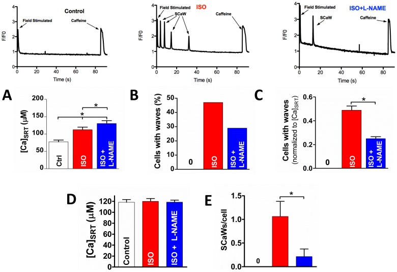
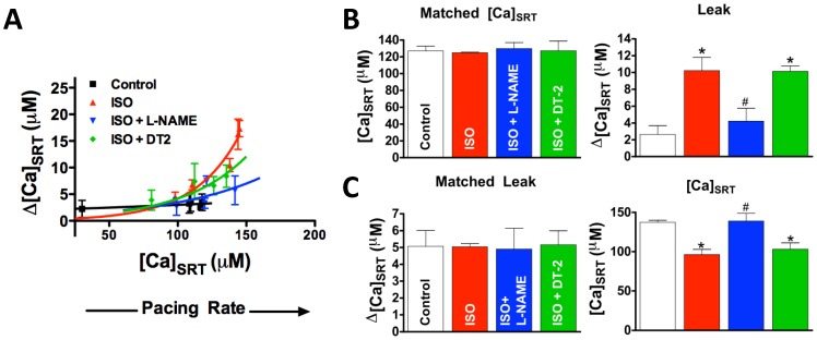
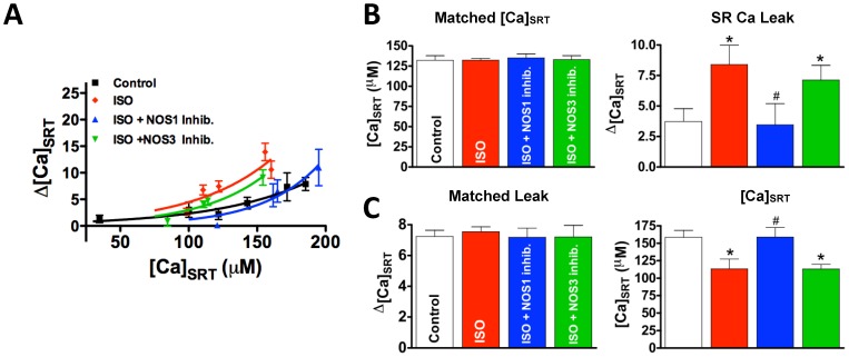
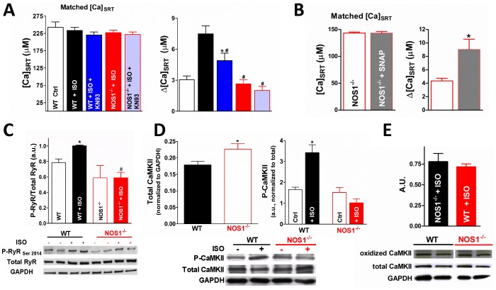
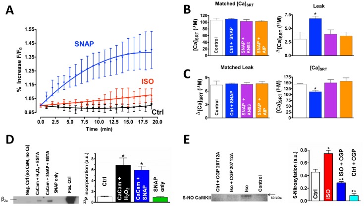
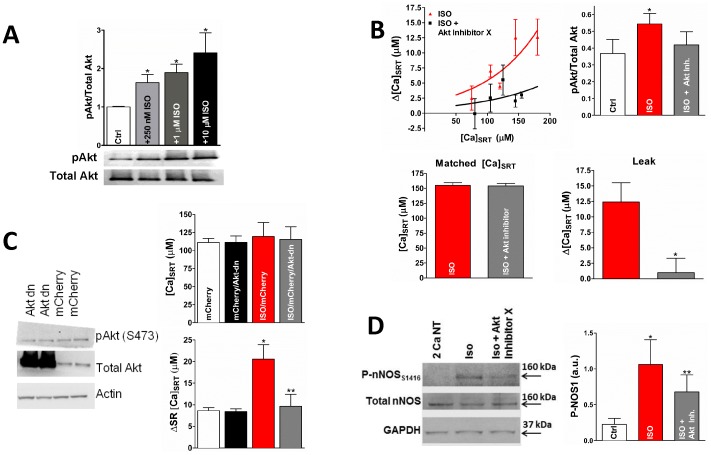
Spontaneous calcium waves in cardiac myocytes are caused by diastolic sarcoplasmic reticulum release (SR Ca(2+) leak) through ryanodine receptors. Beta-adrenergic (β-AR) tone is known to increase this leak through the activation of Ca-calmodulin-dependent protein kinase (CaMKII) and the subsequent phosphorylation of the ryanodine receptor. When β-AR drive is chronic, as observed in heart failure, this CaMKII-dependent effect is exaggerated and becomes potentially arrhythmogenic. Recent evidence has indicated that CaMKII activation can be regulated by cellular oxidizing agents, such as reactive oxygen species. Here, we investigate how the cellular second messenger, nitric oxide, mediates CaMKII activity downstream of the adrenergic signaling cascade and promotes the generation of arrhythmogenic spontaneous Ca(2+) waves in intact cardiomyocytes. Both SCaWs and SR Ca(2+) leak were measured in intact rabbit and mouse ventricular myocytes loaded with the Ca-dependent fluorescent dye, fluo-4. CaMKII activity in vitro and immunoblotting for phosphorylated residues on CaMKII, nitric oxide synthase, and Akt were measured to confirm activity of these enzymes as part of the adrenergic cascade. We demonstrate that stimulation of the β-AR pathway by isoproterenol increased the CaMKII-dependent SR Ca(2+) leak. This increased leak was prevented by inhibition of nitric oxide synthase 1 but not nitric oxide synthase 3. In ventricular myocytes isolated from wild-type mice, isoproterenol stimulation also increased the CaMKII-dependent leak. Critically, in myocytes isolated from nitric oxide synthase 1 knock-out mice this effect is ablated. We show that isoproterenol stimulation leads to an increase in nitric oxide production, and nitric oxide alone is sufficient to activate CaMKII and increase SR Ca(2+) leak. Mechanistically, our data links Akt to nitric oxide synthase 1 activation downstream of β-AR stimulation. Collectively, this evidence supports the hypothesis that CaMKII is regulated by nitric oxide as part of the adrenergic cascade leading to arrhythmogenesis.


Conflict of interest statement

Competing Interests: The authors have declared that no competing interests exist.

Figures

Figure 1
Figure 1. Inhibition of NOS attenuates SCaW formation in ISO treated myocytes.
A) Average [Ca]SRT (n = 34–40) for each treatment (raw data at the top). B) Percentage of myocytes showing at least one SCaW. C) Data in B, normalized to myocyte [Ca]SRT. D&E) [Ca]SRT matched data (D) and the average number of SCaWs exhibited (E, n = 13–15). t-test, *p<0.05).
Figure 2
Figure 2. ISO-dependent leak is attenuated by NOS inhibitor, L-NAME.
A) The leak-dependent shift of Ca2+ from the cytosol to the SR. Each point represents a loading protocol (from low to high [Ca]SRT; resting, 1 field stimulation, 0.25 Hz, 0.5 Hz and 1 Hz stimulation, respectively). B) The SR Ca2+ leak (right) in [Ca]SRT matched data (left, n = 10–14). C) The [Ca]SRT (right) needed to induce the same SR Ca2+ leak (left) in leak matched data (left, n = 11–17). *Statistically different from control, #different from ISO (t-test, p<0.05).
Figure 3
Figure 3. Inhibition of NOS1 but not NOS3 reverses the ISO-dependent increase in SR Ca2+ leak.
A) Leak/load relationship. B) Matched data such that the average [Ca]SRT was the same for all treatments (left) and resultant leaks (right, n = 13–17). C) Data matched such that the average SR Ca2+ leak was the same for all treatments (left) and the [Ca]SRT needed to induce that leak (right, n = 11–19). *different from control, # different from ISO (t-test, p<0.05).
Figure 4
Figure 4. NOS1−/− mice show attenuated CaMKII-dependent leak.
A) Matched data such that [Ca]SRT was the same for all treatments (left) an resultant SR Ca leaks (right, n = 10–22). B) Matched data such that [Ca]SRT was the same in NOS1−/− and NOS1−/−+SNAP (left) and the resultant SR Ca leaks (right), demonstrates that SNAP restores the leak/load relationship in NOS1−/− myocytes. C) Summary data (top, n = 4 hearts each) and representative immunoblot (bottom) of phosphorylated RyR at CaMKII-specific residue, Ser2814, and total RyR expression in WT and NOS1−/− heart lysates. D) Western blots showing total CaMKII normalized to GAPDH (left) and CaMKII phosphorylated at T286 (right, n = 5) in WT and NOS1−/− hearts. Representative blot showing at bottom. E) Summary data (top) and representative immunoblot (bottom) of oxidize CaMKII after ISO stimulation in WT and NOS1−/− heart lysates. *Statistically different from control, # different from WT+ISO (t-test, p<0.05).
Figure 5
Figure 5. NO increases CaMKII-dependent SR Ca2+ leak.
A) NO-dependent DAF-2 fluorescence (n = 6). Spearman correlation = 1.0 for SNAP, 0.9 for ISO, and −0.05 for control. B) SNAP-dependent SR Ca2+ leak. The SR Ca2+ leak (right) in [Ca]SRT matched data (left, n = 9–13). C) Data was matched such that leak was the same (left) with the [Ca]SRT needed to induced that leak shown on the right (n = 12–17). D) Purified CaMKII pre-activated with 200 µM Ca2+ and CaM. H2O2 (Lane 2) or 500 µM SNAP (Lane 3) was added followed by EGTA. ATP32 was added along with purified β2a L-type Ca2+ channel subunit on nickel beads. Incorporation of P32 was measured as an indicator of Ca-independent sustained kinase activity. Lane 1 is CaMKII without Ca2+, CaM, or ATP; Lane 4 is CaMKII without Ca2+, CaM, or ATP plus the addition of SNAP (500 µM) alone. Lane 5 is P32 incorporation in the continued presence of Ca2+ and CaM. E) Cardiac myocytes were field stimulated at 0.5 Hz under the indicated conditions. CaMKII was then immunoprecipitated from cellular homogenates which were then blotted with antibody to S-NO. *different from ISO, **different from both ISO and control (t-test, p<0.05).
Figure 6
Figure 6. Akt Activates NOS.
A) Western blot indicating that ISO increases Akt phosphorylation at S473 in a dose-dependent manner in isolated rabbit cells. B) ISO-dependent increase in p-Akt is blunted by Akt-Inhibitor X (top, right, * different from control, ** different from ISO, paired t-test, p<0.05). Cells were treated with ISO or ISO+Akt Inhibitor X. Akt Inhibitor X decreased the SR Ca leak and the activation of Akt (top left and bottom). C) Down-regulated Akt expression (plus the constitutively expressed Akt) increased the total Akt over the constitutive expression alone (top). Akt-dn decreased ISO-dependent SR Ca leak when data was selected to give the same average [Ca]SRT (bottom). D) NOS1 phosphorylation at Akt phosphorylation site S1416, representative immunoblot (left) and summary data (left). (* different from control, ** different from ISO, paired t-test, p<0.05).

References

    1. Bers DM, Ziolo MT (2001) When is cAMP not cAMP? Effects of compartmentalization. CircRes 89: 373–375. - PubMed
    1. Bers DM (2001) Excitation-Contraction Coupling and Cardiac Contractile Force: Kluwer Academic Publishers.
    1. Dibb KM, Graham HK, Venetucci LA, Eisner DA, Trafford AW (2007) Analysis of cellular calcium fluxes in cardiac muscle to understand calcium homeostasis in the heart. Cell Calcium 42: 503–512. - PubMed
    1. Wang H, Kohr MJ, Traynham CJ, Wheeler DG, Janssen PM, et al. (2008) Neuronal nitric oxide synthase signaling within cardiac myocytes targets phospholamban. Am J Physiol Cell Physiol 294: C1566–1575. - PMC - PubMed
    1. Curran J, Brown KH, Santiago DJ, Pogwizd S, Bers DM, et al. (2010) Spontaneous Ca waves in ventricular myocytes from failing hearts depend on Ca(2+)-calmodulin-dependent protein kinase II. J Mol Cell Cardiol - PMC - PubMed

Publication types

MeSH terms