. 2014 Feb 3;9(2):e87922.
doi: 10.1371/journal.pone.0087922. eCollection 2014.

Concentration-dependent exchange of replication protein A on single-stranded DNA revealed by single-molecule imaging

Affiliations

Concentration-dependent exchange of replication protein A on single-stranded DNA revealed by single-molecule imaging

Bryan Gibb et al. PLoS One. .

Abstract

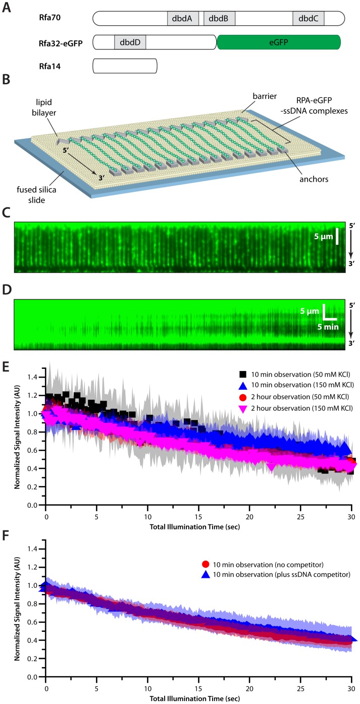
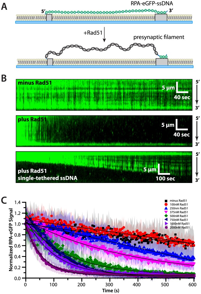
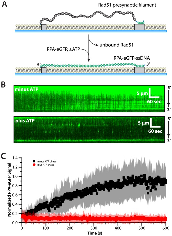
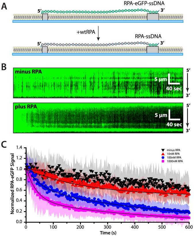
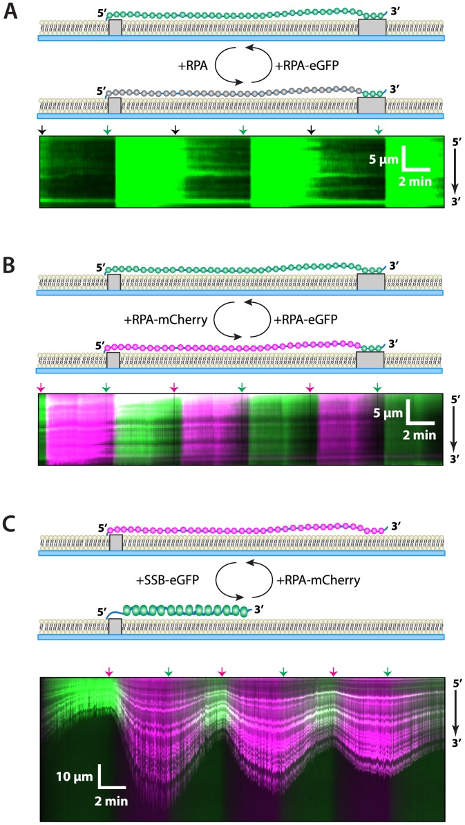
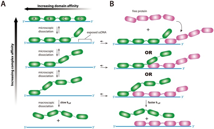
Replication protein A (RPA) is a ubiquitous eukaryotic single-stranded DNA (ssDNA) binding protein necessary for all aspects of DNA metabolism involving an ssDNA intermediate, including DNA replication, repair, recombination, DNA damage response and checkpoint activation, and telomere maintenance. The role of RPA in most of these reactions is to protect the ssDNA until it can be delivered to downstream enzymes. Therefore a crucial feature of RPA is that it must bind very tightly to ssDNA, but must also be easily displaced from ssDNA to allow other proteins to gain access to the substrate. Here we use total internal reflection fluorescence microscopy and nanofabricated DNA curtains to visualize the behavior of Saccharomyces cerevisiae RPA on individual strands of ssDNA in real-time. Our results show that RPA remains bound to ssDNA for long periods of time when free protein is absent from solution. In contrast, RPA rapidly dissociates from ssDNA when free RPA is present in solution allowing rapid exchange between the free and bound states. In addition, the S. cerevisiae DNA recombinase Rad51 and E. coli single-stranded binding protein (SSB) also promote removal of RPA from ssDNA. These results reveal an unanticipated exchange between bound and free RPA suggesting a binding mechanism that can confer exceptionally slow off rates, yet also enables rapid displacement through a direct exchange mechanism that is reliant upon the presence of free ssDNA-binding proteins in solution. Our results indicate that RPA undergoes constant microscopic dissociation under all conditions, but this is only manifested as macroscopic dissociation (i.e. exchange) when free proteins are present in solution, and this effect is due to mass action. We propose that the dissociation of RPA from ssDNA involves a partially dissociated intermediate, which exposes a small section of ssDNA allowing other proteins to access to the DNA.


Conflict of interest statement

Competing Interests: The authors have declared that no competing interests exist.

Figures

Figure 1
Figure 1. Single-stranded DNA curtain assay for RPA binding.
(A) Schematic illustration of S. cerevisiae RPA showing the location of the four primary DNA-binding domains (dbdA-D) and the location of the eGFP tag at the C-terminus of RPA32. (B) Overview of RPA-ssDNA curtains showing the nanofabricated patterns on the surface of a fused silica microscope slide. All of the ssDNA molecules are anchored with their 5′ ends aligned along the leading edges of zig-zag shaped chromium (Cr) barriers , and their 3′ ends anchored through nonspecific adsorption to the exposed Cr pentagons, as depicted . (C) Wide-field TIRF microscopy image of an ssDNA-curtain bound by RPA-eGFP. The 5′ to 3′ orientation of the ssDNA is indicated. Also see Video S1. (D) Kymograph showing a single RPA-eGFP/ssDNA complex with 100-msec images collected at 24-second intervals over a period of 2 hours. (E) Loss of RPA-eGFP signal is due to photo-bleaching. (F) Dissociation of RPA-eGFP is not accelerated in the presence of 1 µM competitor ssDNA. For both (E) and (F) intensity measurements for RPA-eGFP/ssDNA complexes viewed at 2-second intervals for a period of 10 minutes, or at 24-second intervals over 2 hours, as indicated. The total laser illumination period was the same under both experimental conditions. Each curve represents the normalized average calculated from 11–22 different ssDNA molecules collected at 50 or 150 mM KCl, and shaded regions correspond to the standard deviation for each data set.
Figure 2
Figure 2. RPA-eGFP can be rapidly replaced from ssDNA by Rad51.
(A) Schematic illustrating the predicted outcome for an ssDNA curtain experiment (side view) where RPA-eGFP is replaced by unlabeled Rad51. The loss of fluorescence as RPA-eGFP is displaced by Rad51 also coincides with an increase in the length of the ssDNA, which causes an increase in the transverse fluctuations of the ssDNA molecules. (B) The upper panel shows a kymograph of RPA-eGFP bound to ssDNA over time in the absence of Rad51, and the middle panel shows how RPA-eGFP is rapidly displaced from the ssDNA upon injection of 750 nM unlabeled Rad51 with 2.5 mM ATP. Also see Video S2. The lower panel shows an example of a single-tethered ssDNA molecule, which illustrates how Rad51 binding coincides with displacement of RPA-eGFP and extension of the ssDNA. This single-tethered measurement was made using 650 nM Rad51 and 1 mM ATP. (C) RPA-eGFP signal versus time collected at different concentrations of Rad51 (as indicated) in the presence of 2.5 mM ATP in buffer containing 50 mM KCl. Each curve represents the normalized average calculated from 11 to 70 different ssDNA molecules. Shaded regions correspond to the standard deviation for each data set. The data were fit to single exponential decays (solid lines), and loss of signal reflects a combination of photo-bleaching (as reflected in the minus Rad51 control), Rad51-induced dissociation of RPA-eGFP, and corresponding extension of the ssDNA, which causes the time-averaged position of the molecules to move further away from the surface.
Figure 3
Figure 3. ATP prevents dissociation of Rad51 from ssDNA even when free RPA is present.
(A) Experimental schematic illustrating the how replacement of wild-type, dark Rad51 with RPA-eGFP can be used to monitor disassembly of the presynaptic complex on double-tethered ssDNA curtains. (B) Examples of kymographs showing examples of wild-type Rad51 presynaptic complex disassembly reactions on single ssDNA molecules in the absence (upper panel) and presence (lower panel) of 2.5 mM ATP and 1 nM RPA-eGFP at 50 mM KCl. (C) RPA-eGFP fluorescence signal versus time during the Rad51 disassembly reactions. Each curve represents the normalized average calculated from 15 to 20 different ssDNA molecules, and shaded regions correspond to the standard deviation for each data set. When ATP is omitted from the chase buffer, the RPA-eGFP signal increases, reflecting the dissociation of Rad51 from the ssDNA. RPA-eGFP fails to bind to the ssDNA when 2.5 mM ATP is present in the chase buffer, indicating that Rad51 does not dissociate from the ssDNA.
Figure 4
Figure 4. Concentration-dependent exchange of ssDNA-bound RPA.
(A) Schematic illustrating the predicted outcome for an ssDNA curtain experiment (side view) where RPA-eGFP is replaced by unlabeled RPA. (B) The upper panel shows a kymograph of RPA-eGFP bound to ssDNA over time in the absence of free, unlabeled RPA, and the middle panel shows how RPA-eGFP is rapidly replaced upon injection of 1000 nM unlabeled RPA at 50 mM KCl. (C) RPA-eGFP signal versus time collected after the injection of different concentrations of unlabeled RPA (as indicated). Each curve represents the normalized average calculated from 15 to 33 different ssDNA molecules, and the shaded regions correspond to the standard deviation for each data set. The RPA chase data were fit to double exponential decays (solid lines), and loss of signal reflects a combination of photo-bleaching (as reflected in the minus RPA control), and unlabeled RPA-induced dissociation of RPA-eGFP, which increases at higher concentrations of free RPA. The minus RPA reference data set is that same as is shown in Figure 2C.
Figure 5
Figure 5. Two-color experiment showing exchange of bound and free RPA.
Schematic depictions, kymographs, and graphs of exchange experiments conducted with alternating injections of (A) RPA-eGFP and dark, wild-type RPA, (B) RPA-eGFP and RPA-mCherry, or (C) RPA-mCherry (10 nM) and E. coli SSB-eGFP (40 nM). All reactions used buffer containing 150 mM KCl. Arrowheads placed above each kymograph indicate the time point of the protein injections, and are color-coded black, green or magenta to indicate dark protein, eGFP-tagged protein, or mCherry-tagged protein, respectively. The experiments in (A) and (B) used double-tethered ssDNA curtains, whereas the experiment in (C) used single-tethered ssDNA curtains to allow for the ssDNA compaction that accompanies the binding of SSB, as well as the corresponding extension that takes place when SSB is replaced by RPA. Also see Video S3.
Figure 6
Figure 6. Hypothetical model for exchange-dependent dissociation of RPA from ssDNA.
(A) Schematic illustration building on a previously proposed mechanism for binding and dissociation of RPA from ssDNA , and incorporating the concept of microscopic dissociation as a means of driving concentration dependent protein-exchange. During binding each of the four different DNA-binding domains (A to D) sequentially associates with the ssDNA. Intermediates involving submicroscopic dissociation of a subset of the DBDs still retain contact with the ssDNA and cannot macroscopically dissociate into solution, and in the absence of free protein each submicroscopic dissociation step is rapidly reversible. (B) When free ssDNA-binding proteins are present in solution, submicroscopic dissociation of any subset of the RPA DBDs will expose a small patch of ssDNA, providing the opportunity for the new proteins (shown in magenta) to bind the ssDNA. The presence of the newly bound protein will restrict re-association of the microscopically dissociated RPA DBD, thereby promoting macroscopic dissociation into solution of the original RPA molecule (shown in green).

References

    1. Broderick S, Rehmet K, Concannon C, Nasheuer HP (2010) Eukaryotic single-stranded DNA binding proteins: central factors in genome stability. Subcell Biochem 50: 143–163. - PubMed
    1. Oakley GG, Patrick SM (2010) Replication protein A: directing traffic at the intersection of replication and repair. Front Biosci 15: 883–900. - PMC - PubMed
    1. Wold MS (1997) Replication protein A: a heterotrimeric, single-stranded DNA-binding protein required for eukaryotic DNA metabolism. Annu Rev Biochem 66: 61–92. - PubMed
    1. Fanning E, Klimovich V, Nager AR (2006) A dynamic model for replication protein A (RPA) function in DNA processing pathways. Nucleic Acids Res 34: 4126–4137. - PMC - PubMed
    1. Fan J, Pavletich NP (2012) Structure and conformational change of a replication protein A heterotrimer bound to ssDNA. Genes Dev 26: 2337–2347. - PMC - PubMed

Publication types

MeSH terms