. 2014 Feb 3;9(2):e88111.
doi: 10.1371/journal.pone.0088111. eCollection 2014.

Shared gene structures and clusters of mutually exclusive spliced exons within the metazoan muscle myosin heavy chain genes

Affiliations

Shared gene structures and clusters of mutually exclusive spliced exons within the metazoan muscle myosin heavy chain genes

Martin Kollmar et al. PLoS One. .

Abstract

Multicellular animals possess two to three different types of muscle tissues. Striated muscles have considerable ultrastructural similarity and contain a core set of proteins including the muscle myosin heavy chain (Mhc) protein. The ATPase activity of this myosin motor protein largely dictates muscle performance at the molecular level. Two different solutions to adjusting myosin properties to different muscle subtypes have been identified so far: Vertebrates and nematodes contain many independent differentially expressed Mhc genes while arthropods have single Mhc genes with clusters of mutually exclusive spliced exons (MXEs). The availability of hundreds of metazoan genomes now allowed us to study whether the ancient bilateria already contained MXEs, how MXE complexity subsequently evolved, and whether additional scenarios to control contractile properties in different muscles could be proposed, By reconstructing the Mhc genes from 116 metazoans we showed that all intron positions within the motor domain coding regions are conserved in all bilateria analysed. The last common ancestor of the bilateria already contained a cluster of MXEs coding for part of the loop-2 actin-binding sequence. Subsequently the protostomes and later the arthropods gained many further clusters while MXEs got completely lost independently in several branches (vertebrates and nematodes) and species (for example the annelid Helobdella robusta and the salmon louse Lepeophtheirus salmonis). Several bilateria have been found to encode multiple Mhc genes that might all or in part contain clusters of MXEs. Notable examples are a cluster of six tandemly arrayed Mhc genes, of which two contain MXEs, in the owl limpet Lottia gigantea and four Mhc genes with three encoding MXEs in the predatory mite Metaseiulus occidentalis. Our analysis showed that similar solutions to provide different myosin isoforms (multiple genes or clusters of MXEs or both) have independently been developed several times within bilaterian evolution.


Conflict of interest statement

Competing Interests: The authors have declared that no competing interests exist.

Figures

Figure 1
Figure 1. The unrooted phylogenetic split network was generated with SplitsTree using the NeighborNet method.
The network presents alternative splits in the evolution of the muscle myosin heavy chain (Mhc) proteins. The Schizosaccharomyces non-muscle Mhc proteins have been used as outgroup. The phylogenetic trees based on the same data using three different methods are shown in Figure S1.
Figure 2
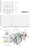
Figure 2. Conserved intron positions and location of MXE encoded regions.
A) The gene structure alignment was generated with Genepainter by mapping intron positions obtained from the gene structure reconstructions onto the protein multiple sequence alignment. Genepainter requires intron positions not only conserved at the amino-acid level but also at the nucleotide level (codons might be split differently). Hyphens “-” represent coding regions and vertical bars “|” denote intron positions. Common intron positions in the gene structure alignment are conserved down to the nucleotide level. Conserved clusters of MXEs are colour coded and numbered from N- to C-terminus (see legend). The same colour coding and numbering scheme will be used throughout this analysis for all MXEs. Some branch names are given for better orientation. B) The structure of the motor domain of the non-muscle class-II myosin of Dictyostelium discoideum has been used to highlight the regions encoded by alternatively spliced exons. For colouring the regions encoded by MXEs the same colours have been used as for the gene structures in A). The clusters of MXEs not described so far code for the light-green (cluster-3) and the dark-brown (cluster-4) part of the structure.
Figure 3
Figure 3. The ctenophore Mnemiopsis leidyi contains two muscle Mhc genes (left side) of which one contains a cluster of MXEs.
Exons and introns are represented as dark- and light-grey bars, respectively, MXEs are shown in colour. The opacity of the colour of the 3′ of the alternative exons corresponds to the alignment score of the alternative exon to the original one (5′ exon). A legend is given explaining the colour coding of features within the gene structure schemes. On the right side, the structural region covered by the MXEs is shown mapped onto the crystal structure of the motor domain of the Dictyostelium discoideum non-muscle myosin protein .
Figure 4
Figure 4. Examples of Platyhelminthes Mhc genes.
A) The freshwater planarian Schmidtea mediterranea contains three Mhc genes. In all gene structure schemes exons and introns are represented as dark- and light-grey bars, MXEs are shown in colour. The opacity of the colour of the 3′ of the alternative exons corresponds to the alignment score of the alternative exon to the original one (5′ exon). B) Sequence alignment of the myosin proteins of the analysed Platyhelminthes around the loop-2 region. The part of loop-2, which is encoded by MXEs, is indicated. The sequences of the 5′ exons are very similar across the Platyhelminthes, as are the 3′ exons, implying that the ancestor of the Platyhelminthes already contained this cluster of MXEs.
Figure 5
Figure 5. Examples of annelid and mollusc Mhc genes, and location of the MXE coding regions within the motor domain.
A) The annelid Helobdella robusta contains two Mhc genes without any clusters of MXEs, while the annelid Capitella teleta contains one Mhc gene with many clusters of MXEs and three differentially included exons at the C-terminus. B) The structural regions covered by MXEs present in lophotrochozoans are shown mapped onto the crystal structure of the motor domain of the Dictyostelium discoideum non-muscle myosin protein . C) Examples of representative mollusc Mhc genes showing the divergence in MXE clusters in the respective subphyla. D) Gene structures of the muscle Mhc genes in the owl limpet Lottia gigantea. The scheme at the bottom shows the genomic region of the cluster of Mhc genes including the Mhc8 gene that encodes only part of the coiled-coil tail region. Reading direction is designated by arrows. Colours of exons in the Mhc gene cluster represent exons coding for a similar part of the protein. In all gene structure schemes exons and introns are represented as dark- and light-grey bars, MXEs are shown in colour. The opacity of the colour of the 3′ of the alternative exons corresponds to the alignment score of the alternative exon to the original one (5′ exon). The vertical red line in the genomic region scheme at the bottom represents a region of unknown sequence (“N”s). The complete list of lophotrochozoan Mhc genes is shown in Figure S2.
Figure 6
Figure 6. The schemes represent examples of Mhc genes in Chelicerata.
Metaseiulus occidentalis contains four Mhc genes of which the ones with MXE clusters are arranged as tandem array of gene duplicates (Mhc3, Mhc4 and Mhc5). Parasteatoda tepidariorum and Centruroides sculpturatus both contain two Mhc genes. The complete list of Chelicerata Mhc genes is shown in Figure S2. Exons and introns are represented as dark- and light-grey bars, MXEs are shown in colour. The opacity of the colour of the 3′ of the alternative exons corresponds to the alignment score of the alternative exon to the original one (5′ exon).
Figure 7
Figure 7. Examples of Mhc genes of Palaeoptera, Coleoptera and Paraneoptera.
The examples have been chosen because of the unusual combinations of clusters of MXEs or because of unusual high or low numbers of MXEs within clusters. The complete list of arthropod Mhc genes is shown in Figure S2. Exons and introns are represented as dark- and light-grey bars, MXEs are shown in colour. The opacity of the colour of the 3′ of the alternative exons corresponds to the alignment score of the alternative exon to the original one (5′ exon).
Figure 8
Figure 8. Schematic drawing of the evolution of the clusters of MXEs within eumetazoan Mhc genes.
The only Ctenophora sequenced, Mnemiopsis leidyi, contains a cluster of MXEs that does not correspond to any other known cluster and has therefore been named cluster-0. The tree is shown as schematic tree representing known phylogenetic relationships to which MXE cluster loss and gain events were plotted. MXE clusters were regarded as gained in the last common ancestor of the branch, which contains species encoding these clusters. According to this scheme, five clusters have evolved in the last common ancestor of the Protostomia, and a set of three clusters later at the onset of the arthropods. There are many branches and species that completely lost all clusters of MXEs in their Mhc genes. Coloured boxes represent MXE cluster gain events (tree view, left side) and their presence within a certain branch (table, right side). Crossed boxes denote MXE cluster loss events. MXEs in light-colour symbolize clusters of MXEs that were supposed to be present but could not be approved because of genome assembly gaps (Figure S2).

Similar articles

Cited by

References

    1. Pohl M, Bortfeldt RH, Grützmann K, Schuster S (2013) Alternative splicing of mutually exclusive exons–a review. BioSystems 114: 31–38 10.1016/j.biosystems.2013.07.003 - DOI - PubMed
    1. Hatje K, Kollmar M (2013) Expansion of the mutually exclusive spliced exome in Drosophila. Nat Commun 4: 2460 10.1038/ncomms3460 - DOI - PubMed
    1. Graveley BR (2005) Mutually Exclusive Splicing of the Insect Dscam Pre-mRNA Directed by Competing Intronic RNA Secondary Structures. Cell 123: 65–73 10.1016/j.cell.2005.07.028 - DOI - PMC - PubMed
    1. Odronitz F, Kollmar M (2008) Comparative genomic analysis of the arthropod muscle myosin heavy chain genes allows ancestral gene reconstruction and reveals a new type of “partially” processed pseudogene. BMC Mol Biol 9: 21 10.1186/1471-2199-9-21 - DOI - PMC - PubMed
    1. Pillmann H, Hatje K, Odronitz F, Hammesfahr B, Kollmar M (2011) Predicting mutually exclusive spliced exons based on exon length, splice site and reading frame conservation, and exon sequence homology. BMC Bioinformatics 12: 270 10.1186/1471-2105-12-270 - DOI - PMC - PubMed

Publication types

Substances