. 2014 Jan 30;8(1):e2664.
doi: 10.1371/journal.pntd.0002664. eCollection 2014.

Leptospira Interrogans induces fibrosis in the mouse kidney through Inos-dependent, TLR- and NLR-independent signaling pathways

Affiliations

Leptospira Interrogans induces fibrosis in the mouse kidney through Inos-dependent, TLR- and NLR-independent signaling pathways

Martine Fanton d'Andon et al. PLoS Negl Trop Dis. .

Erratum in

  • PLoS Negl Trop Dis. 2014 Apr;8(4):e2802

Abstract

Background: Leptospira (L.) interrogans are bacteria responsible for a worldwide reemerging zoonosis. Rodents carry L. interrogans asymptomatically in their kidneys and excrete bacteria in the urine, contaminating the environment. Humans get infected through skin contact and develop a mild or severe leptospirosis that may lead to renal failure and fibrosis. L. interrogans provoke an interstitial nephritis, but the induction of fibrosis caused by L. interrogans has not been studied in murine models. Innate immune receptors from the TLR and NLR families have recently been shown to play a role in the development and progression of tissue fibrosis in the lung, liver and kidneys under different pathophysiological situations. We recently showed that TLR2, TLR4, and NLRP3 receptors were crucial in the defense against leptospirosis. Moreover, infection of a human cell line with L. interrogans was shown to induce TLR2-dependent production of fibronectin, a component of the extracellular matrix. Therefore, we thought to assess the presence of renal fibrosis in L. interrogans infected mice and to analyze the contribution of some innate immune pathways in this process.

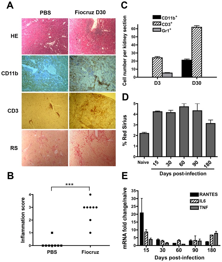
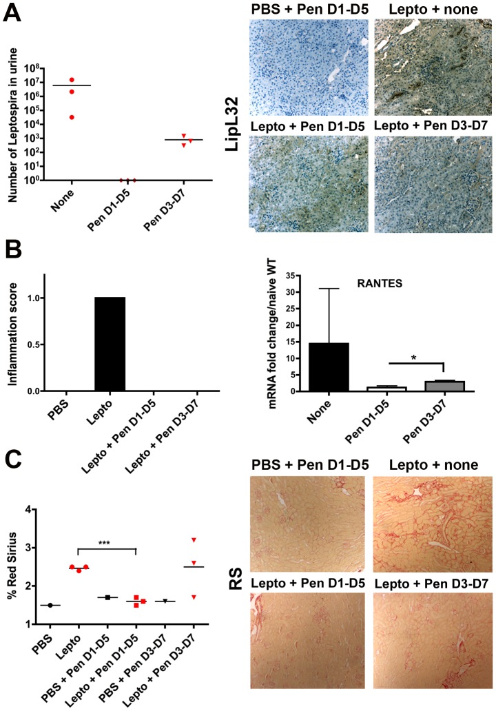
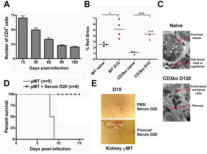
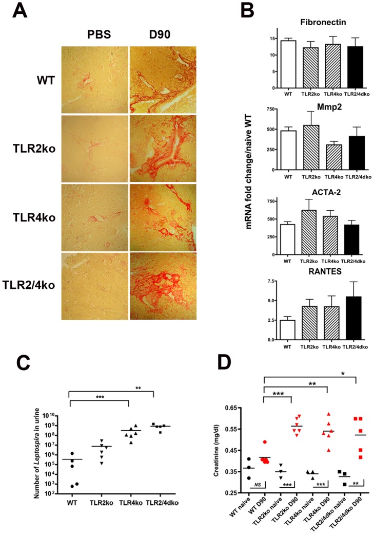
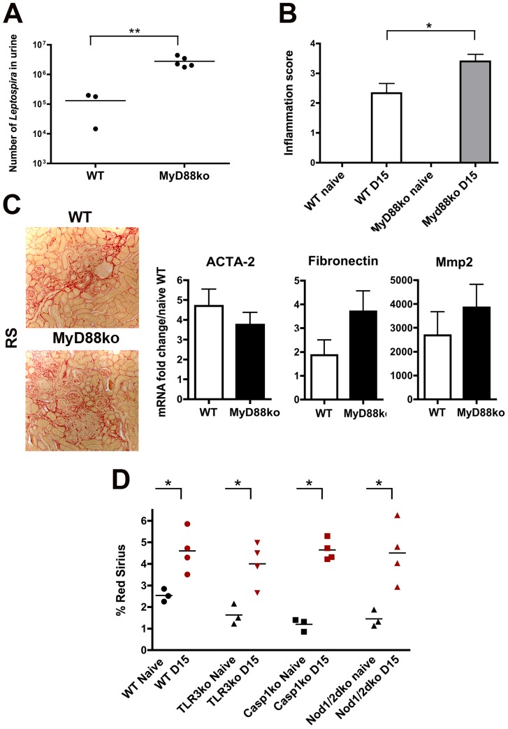
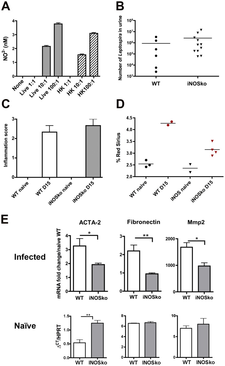
Methodology/principal findings: Here, we characterized by immunohistochemical studies and quantitative real-time PCR, a model of Leptospira-infected C57BL/6J mice, with chronic carriage of L. interrogans inducing mild renal fibrosis. Using various strains of transgenic mice, we determined that the renal infiltrates of T cells and, unexpectedly, TLR and NLR receptors, are not required to generate Leptospira-induced renal fibrosis. We also show that the iNOS enzyme, known to play a role in Leptospira-induced interstitial nephritis, also plays a role in the induction of renal fibrosis.

Conclusion/significance: To our knowledge, this work provides the first experimental murine model of sustained renal fibrosis induced by a chronic bacterial infection that may be peculiar, since it does not rely on TLR or NLR receptors. This model may prove useful to test future therapeutic strategies to combat Leptospira-induced renal lesions.


Conflict of interest statement

The authors declared that they have no competing interests.

Figures

Figure 1
Figure 1. Leptospira infection triggers inflammation and fibrosis in the mouse kidney.
(A) Light microscopy of nodular infiltrates stained with hematoxylin-eosin (HE), infiltrating CD11b+ macrophages and CD3+ T cells and collagen deposition stained with Red Sirius (RS) in kidneys from C57BL/6J mice 30 days (D30) after the inoculation of 2×10⁸ L. interrogans strain Fiocruz. As controls, mice were injected with PBS. Magnification, ×100. (B) Score of kidney inflammation of interstitial nodular infiltrates per surface areas from five different renal tissue sections in control (PBS) and L. interrogans strain Fiocruz infected mice (n = 8 per group). (C) Quantification of the number of CD11b+ macrophages, Gr1+ neutrophils and CD3+ T Lymphocytes per surface area in kidneys from day-3 (D3) and day-30 post-infected mice. (D) Fibrosis quantification by Red Sirius morphometry, expressed as percent of surface area and (E) Inflammation evaluation by mRNA expression of proinflammatory mediators in kidneys of 10 infected mice sacrificed at different time points. Values are means ± SD of counts (C and D) and mRNA quantification (E) from 5 different tissue sections from n = 2 separate mice in each group tested. ***P<0.001.
Figure 2
Figure 2. Effects of antibiotic treatment on Leptospira-infected mice.
C57BL/6J mice were infected with 107 L. interrogans serovar Manilae (n = 3) or PBS (n = 1) and injected (IP) daily for 5 days with penicillin G (Pen) from day-1 p.i. until day-5 (D1–D5) or from day-3 p.i. until day-7 (D3–D7). Thereafter, mice were sacrificed at day-24 p.i. (A) Leptospiral loads in 100 µl urine determined by qRT-PCR, and imaging of the LipL32 leptospiral major antigen immunostaining in kidney sections. (B) Inflammation score (left panel) and pro-inflammatory RANTES mRNA expression (right panel). (C) Quantification (left panel) and microscopy (right panels) of fibrosis by Red Sirius staining. Values are counts (The bars represent the mean value in each group) or means of mRNA quantification ± SD from n = 3 mice per group and are from one representative experiment of two. *P<0.05; ***P<0.001 between groups.
Figure 3
Figure 3. T and B cells are not involved in Leptospira-induced renal fibrosis.
(A) Quantification of the number of infiltrating CD3+ cells in Leptospira-infected kidneys from WT C57BL/6J mice at different time points post p.i.. Values are means ± SD from five kidney sections per surface area from (n = 2) mice at each different time point tested. (B) Percentage of Red Sirius labeling per surface area in kidney sections from WT and CD3ko mice at day-15 p.i. (D15) and in naive mice. The bars represent the mean value in each group. (C) Electron microscopy (magnification ×100) of kidneys sections from a naïve mouse and CD3ko mouse at 4 months p.i. (D120) showing fibrosis. (D) Survival curves and (E) images of renal Red Sirius staining from B cell deficient naïve μMT mice or infected with 107 L. interrogans strain Fiocruz, rescued by immune serum from Leptospira-infected WT mice at day-15 p.i. (D15). *P<0.05; ***P<0.001 between groups.
Figure 4
Figure 4. TLR2 and TLR4 are not involved in Leptospira-induced fibrosis.
(A–D) Renal fibrosis, levels of inflammatory mediators, bacterial loads, and serum creatinine levels in WT or TLR2 and/or TLR4 deficient mice infected with 2×10⁶ L. interrogans Fiocruz at 3 months p.i. (D90) (A) Red Sirius staining. (B) Fibrosis and inflammation evaluation by mRNA expression of fibronectin, Mmp2, ACTA-2 and RANTES, in kidneys of WT, TLR2ko, TLR4ko and TLR2/4dko mice at 3 months p.i. Values are means ± SD from n = 5 mice per genotype group. No statistical difference between genotypes was found for the different markers by One-Way Anova. (C) Bacterial loads in 100 µl of urine from mice at 3 months p.i. The bars represent the mean values in each group tested. (D) Serum creatinine levels in naïve and Leptospira-infected mice at 3 months p.i. *P<0.05; **P<0.01; ***P<0.001 between groups.
Figure 5
Figure 5. TLRs and NLRs are not involved in Leptospira-induced fibrosis.
WT and Myd88ko mice infected with 2×10⁶ L. interrogans Fiocruz were sacrificed at 15 days p.i. (A) Bacterial loads in 100 µl of urine. (B) Inflammation score in kidneys. (C) Red Sirius staining (C, left panel) and mRNA expression of ACTA-2, fibronectin and Mmp2, in the infected WT and Myd88ko mice kidneys (C, right panel). Data are means ± SD from WT mice (n = 3) and MyD88ko mice (n = 5). The images in C are representative of three separate experiments. (D) Percentage of Red Sirius staining in kidneys from WT mice, TLR3ko, Casp1ko and double Nod1/2ko mice infected with 2×10⁸ L. interrogans Fiocruz and sacrificed at day-15 p.i. (n = 4 per genotype group). Three naïve kidneys of each genotype were used as controls. *P<0.05; **P<0.01 between groups.
Figure 6
Figure 6. iNOS is involved in Leptospira-induced renal fibrosis.
NO2 production in supernatants from bone marrow macrophages derived from C57BL/6J mice, stimulated for 24 h with live (Lepto) or heat-killed (HKLepto) L. interrogans strain Fiocruz at a multiplicity of infection of 1 (1∶1) 10 (10∶1) and 100 (100∶1), as measured by the Griess reaction. Values are means +SD from 3 separate experiments. (B–E) Bacterial loads in urine (B), renal inflammatory scores (C), percentage of Red Sirius staining (D) and mRNA expression of ACTA-2, fibronectin and Mmp2 (E, upper panel) in kidneys from WT and iNOSko mice infected with 2×10⁸ L. interrogans Fiocruz and sacrificed at day-15 p.i.. Values are means ± SD from WT (n = 3) and iNOSko (n = 5) mice. (E, lower panel), comparison of mRNA expression levels of fibrosis markers in kidneys of naïve WT and iNOSko mice, measured as ΔCT compared to HPRT. Values are means ± SD from WT (n = 3) and iNOSko (n = 3) mice. *P<0.05; **P<0.01 between groups.

References

    1. Bharti AR, Nally JE, Ricaldi JN, Matthias MA, Diaz MM, et al. (2003) Leptospirosis: a zoonotic disease of global importance. Lancet Infect Dis 3: 757–771. - PubMed
    1. Mc IW, Montgomery GL (1952) Renal lesions in Leptospira canicola infection in dogs. J Pathol Bacteriol 64: 145–160. - PubMed
    1. Atasoyu EM, Turhan V, Unver S, Evrenkaya TR, Yildirim S (2005) A case of leptospirosis presenting with end-stage renal failure. Nephrol Dial Transplant 20: 2290–2292. - PubMed
    1. Yang CW (2007) Leptospirosis in Taiwan–an underestimated infectious disease. Chang Gung Med J 30: 109–115. - PubMed
    1. Bandeira M, Santos CS, de Azevedo EC, Soares LM, Macedo JO, et al. (2011) Attenuated nephritis in inducible nitric oxide synthase knockout C57BL/6 mice and pulmonary hemorrhage in CB17 SCID and recombination activating gene 1 knockout C57BL/6 mice infected with Leptospira interrogans. Infect Immun 79: 2936–2940. - PMC - PubMed

MeSH terms