. 2014 Jan 30;8(1):e2672.
doi: 10.1371/journal.pntd.0002672. eCollection 2014.

Leptospira interrogans binds to cadherins

Affiliations

Leptospira interrogans binds to cadherins

Karen Evangelista et al. PLoS Negl Trop Dis. .

Abstract

Leptospirosis, caused by pathogenic species of Leptospira, is the most widespread zoonosis and has emerged as a major public health problem worldwide. The adhesion of pathogenic Leptospira to host cells, and to extracellular matrix (ECM) components, is likely to be necessary for the ability of leptospires to penetrate, disseminate and persist in mammalian host tissues. Previous work demonstrated that pathogenic L. interrogans binds to host cells more efficiently than to ECM. Using two independent screening methods, mass spectrometry and protein arrays, members of the cadherin family were identified as potential L. interrogans receptors on mammalian host surfaces. We focused our investigation on vascular endothelial (VE)-cadherin, which is widely expressed on endothelia and is primarily responsible for endothelial cell-cell adhesion. Monolayers of EA.hy926 and HMEC-1 endothelial cells produce VE-cadherin, bind L. interrogans in vitro, and are disrupted upon incubation with the bacteria, which may reflect the endothelial damage seen in vivo. Dose-dependent and saturable binding of L. interrogans to the purified VE-cadherin receptor was demonstrated and pretreatment of purified receptor or endothelial cells with function-blocking antibody against VE-cadherin significantly inhibited bacterial attachment. The contribution of VE-cadherin to leptospiral adherence to host endothelial cell surfaces is biologically significant because VE-cadherin plays an important role in maintaining the barrier properties of the vasculature. Attachment of L. interrogans to the vasculature via VE-cadherin may result in vascular damage, facilitating the escape of the pathogen from the bloodstream into different tissues during disseminated infection, and may contribute to the hemorrhagic manifestations of leptospirosis. This work is first to describe a mammalian cell surface protein as a receptor for L. interrogans.


Conflict of interest statement

The authors have declared that no competing interests exist.

Figures

Figure 1
Figure 1. Leptospira interrogans binds to EA.hy926 endothelial cells more efficiently than to ECM.
Confluent cell layers were left intact, or lifted with EDTA to remove all cells from the ECM without degrading receptors. The cells were pelleted, and both the cells and the ECM were washed in medium without antibiotics prior to the addition of 35S-labeled L. interrogans serovar Copenhageni strain Fiocruz L1–130 at an MOI of 10. After 1 hr at 37°C followed by washing, bound bacteria were quantified. Results are shown as the mean ± standard error of 112 replicates from multiple experiments. By one way ANOVA followed by Tukey's multiple comparison test, binding to lifted cells vs. the intact layer was not significantly different, but for binding to ECM vs. either lifted cells or intact layer, P<0.001.
Figure 2
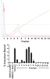
Figure 2. Representative DEAE-sepharose fractionation of EA.hy926 endothelial cell membrane extracts to identify fractions to which L. interrogans bound.
Top panel: FPLC system traces. The red trace denotes absorbance at 254 nm, the blue trace denotes absorbance at 280 nm, the brown trace denotes conductivity. Bottom panel: Binding of L. interrogans to fractions shown in the top panel. The “extract” was the octylglucoside supernatant loaded on to the column. All fractions were diluted 1∶10 in HBSC buffer without detergent prior to dispensing into the wells. After blocking the wells, 3.5×10⁵ 35S-labeled pathogenic L. interrogans serovar Copenhageni was added to wells, and the plate was incubated 1 hour at 37°C, 5% CO2 prior to removal of non-attached bacteria by washing. Buffer with octylglycoside, without or with NaCl, was used as a control for the start and end of the NaCl gradient, respectively.
Figure 3
Figure 3. L. interrogans serovar Copenhageni Fiocruz L1–130 binds to a specific subset of human proteins in an array.
After incubation with the bacteria, the slides were washed and probed with anti-Leptospira protein LipL32, then with fluorophore-conjugated detection reagent. Shown is the human protein array probed with L. interrogans serovar Copenhageni for 3 hours. The complete data set is presented in Supplemental Table S1.
Figure 4
Figure 4. Pathogenic L. interrogans binds to purified cadherins.
Panel A: Purified receptors were diluted to 0.1 µM in HBSC buffer and immobilized in 96 well plates, and non-specific binding sites were blocked. 3.5×10⁵ 35S-labeled pathogenic L. interrogans serovar Copenhageni or saprophytic L. biflexa serovar Patoc was added to wells, and the plate was incubated 1 hour at 37°C, 5% CO2 prior to removal of non-attached bacteria by washing. Bound bacteria were quantified by scintillation counting. Results are shown as the mean ± standard error of 4 replicate determinations of the percent inoculum bound; similar results were obtained from 2 independent experiments. Pathogenic Leptospira bind more efficiently to E-cadherin and VE-cadherin compared to the saprophytic strain (Two-way ANOVA, Bonferroni post-test, P<0.01). Panel B: The purified proteins shown along the x axis were diluted to 0.1 µM in buffer and immobilized in 96 well plates, and attachment of 35S-labeled L. interrogans Copenhageni was assessed by methods described for panel A. Binding of L. interrogans to each cadherin was significantly more efficient than to fibronectin, the positive control (Tukey's multiple comparison test, P<0.05). Panel C: Purified VE-cadherin and E-cadherin were diluted to 0.01, 0.03 and 0.1 µM in HBSC, immobilized in 96-well plates, and then incubated with 3.5×10⁵ radiolabeled bacteria for 1 hr at 37°C, 5% CO2. A dose-dependent attachment of 35S L. interrogans was observed with increasing concentrations of receptors on the wells. Panel D: Increasing numbers of bacteria (1×10³–1×10⁶) were incubated with 0.03 µM VE-cadherin or control super fibronectin immobilized on 96 well plates. The bacterial attachment to VE-cadherin was saturable at 1×10⁴ leptospires. The percent inoculum bound for Panels C and D was determined as previously described in Panel A and results are shown as the mean ± standard error of 4 replicates. For all panels, bovine serum albumin (BSA) served as negative control.
Figure 5
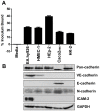
Figure 5. L. interrogans binds to cell monolayers expressing cadherins.
Panel A: Post-confluent endothelial and epithelial monolayers were incubated with 35S labeled L. interrogans serovar Copenhageni strain Fiocruz L1–130 at an MOI of 20. After 1 hr at 37°C followed by washing, bacteria bound to monolayers were quantified and expressed as the mean ± standard error of 8–24 replicates from multiple independent experiments. Panel B: Whole cell lysates were prepared from post-confluent cell monolayers. The cells were collected by scraping followed by treatment with lysis buffer. Fifteen micrograms of the lysate were separated by 10% SDS-PAGE and transferred to an Immobilon membrane. The membranes were probed with antibodies against pan-cadherin (dilution 1∶250), VE-cadherin (dilution 1∶100), E-cadherin (dilution 1∶500), N-cadherin (dilution 1∶200), and ICAM-2 (dilution 1∶2,000). A replicate membrane was probed with anti-GAPDH antibody (dilution 1∶1,000) as a loading control. Molecular sizes of bands were determined by comparing their relative mobilities against the set of standards. Band sizes correspond with expected sizes described by antibody manufacturers.
Figure 6
Figure 6. Anti-VE-cadherin inhibits L. interrogans attachment to the receptor.
Purified VE-cadherin coated on a Linbro plate at 0.1 µM (Panel A) or a post-confluent endothelial cell HMEC-1 monolayer (Panel B) was pretreated with function-blocking anti-VE-cadherin or control antibody IgG2a prior to the addition of 3.5×10⁵ 35S-labeled L. interrogans Copenhageni Fiocruz L1–130. After 1 hour incubation with L. interrogans at 37°C, 5% CO2, the unbound bacteria were removed by washing while the bound bacteria were quantified by scintillation counting. The results are expressed as bacterial attachment relative to untreated wells (without antibody) which was set at 1.0, and are shown as mean ± standard error of 8–24 replicates from multiple independent experiments. A significant decrease in the attachment of L. interrogans was observed when purified receptor was blocked with 20 µg/ml anti-VE-cadherin and when the HMEC-1 monolayer was pretreated with 100 µg/ml anti-VE-cadherin, but no significant differences were seen with the control IgG2a (Tukey's multiple comparison test, P<0.001).

References

    1. Adler B, de la Pena Moctezuma A (2010) Leptospira and leptospirosis. Vetrinary Microbiology 140: 287–296. - PubMed
    1. Ballard SA, Williamson M, Adler B, Vinh T, Faine S (1986) Interactions of virulent and avirulent leptospires with primary cultures of renal epithelial cells. J Med Microbiol 21: 59–67. - PubMed
    1. Ito T, Yanagawa R (1987) Leptospiral attachment to extracellular matrix of mouse fibroblast (L929) cells. Vet Microbiol 15: 89–96. - PubMed
    1. Merien F, Baranton G, Perolat P (1997) Invasion of Vero cells and induction of apoptosis in macrophages by pathogenic Leptospira interrogans are correlated with virulence. Infect Immun 65: 729–738. - PMC - PubMed
    1. Merien F, Truccolo J, Rougier Y, Baranton G, Perolat P (1998) In vivo apoptosis of hepatocytes in guinea pigs infected with Leptospira interrogans serovar icterohaemorrhagiae. FEMS Microbiol Lett 169: 95–102. - PubMed

LinkOut - more resources