Case Reports
. 2014 Jan;29(1):67-70.
doi: 10.5001/omj.2014.15.

A case of rasmussen encephalitis: the differential diagnoses and role of diagnostic imaging

Affiliations
Case Reports

A case of rasmussen encephalitis: the differential diagnoses and role of diagnostic imaging

Binoj Varghese et al. Oman Med J. 2014 Jan.

Abstract

Rasmussen encephalitis is an extremely rare chronic inflammatory neurodegenerative disease affecting a single cerebral hemisphere, causing progressive neurological deterioration and intractable seizures. Imaging plays an important role in diagnosis by demonstrating focal or unihemispheric involvement and excluding other possible causes. Here, we report a case of Rasmussen encephalitis with an update on recent diagnostic criteria and emphasis on differential diagnoses which can be excluded on imaging.

Keywords: Epilepsia partialis continua; Magnetic resonance imaging; Rasmussen encephalitis.


Figures

Figure 1
Figure 1
(a) Axial T2W image (TR: 4224 ms, TE: 99 ms, slice thickness: 4 mm) showing an area of hyperintense signal in left superior frontal region along with widening of cortical sulci on left side; (b) Axial FLAIR image (TR: 7800 ms, TE: 110 ms, slice thickness: 4 mm) showing hyperintense signal in left superior frontal cortex. A small hyperintense focus is also seen in anterior white matter (arrow).
Figure 2
Figure 2
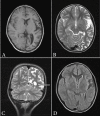
(a) Axial T1-weighted image (TR: 660 ms, TE: 14 ms, slice thickness: 4 mm) showing atrophy of left hemisphere; (b)Axial T2W image (TR: 4224 ms, TE: 99 ms, slice thickness: 4 mm) showing widening of cortical sulci and sylvian fissure on the left side; (c) Coronal T2-weighted image (TR: 5979 ms, TE: 99 ms, slice thickness: 3 mm) demonstrating dilatation of left lateral ventricle (arrow) and widening of cortical sulci; (d) Axial FLAIR image (TR: 7800 ms, TE: 110 ms, slice thickness: 4 mm) showing features of volume loss on the left side.
Figure 3
Figure 3
(a) Axial gradient-echo image (TR: 704 ms, TE: 15 ms, slice thickness: 4 mm) showing no abnormal gyral calcification; (b) Axial T1-weighted image (TR: 660 ms, TE: 14 ms, slice thickness: 4 mm) showing normally developed frontal sinus and mastoid air cells.

References

    1. Rasmussen T, Olszewski J, Lloydsmith D. Focal seizures due to chronic localized encephalitis. Neurology 1958. Jun;8(6):435-445 10.1212/WNL.8.6.435 - DOI - PubMed
    1. Bien CG, Bauer J, Deckwerth TL, Wiendl H, Deckert M, Wiestler OD, et al. Destruction of neurons by cytotoxic T cells: a new pathogenic mechanism in Rasmussen’s encephalitis. Ann Neurol 2002a Mar;51(3):311-318 10.1002/ana.10100 - DOI - PubMed
    1. Bien CG, Granata T, Antozzi C, Cross JH, Dulac O, Kurthen M, et al. Pathogenesis, diagnosis and treatment of Rasmussen encephalitis: a European consensus statement. Brain 2005. Mar;128(Pt 3):454-471 10.1093/brain/awh415 - DOI - PubMed
    1. Rogers SW, Andrews PI, Gahring LC, Whisenand T, Cauley K, Crain B, et al. Autoantibodies to glutamate receptor GluR3 in Rasmussen’s encephalitis. Science 1994. Jul;265(5172):648-651 10.1126/science.8036512 - DOI - PubMed
    1. Bien CG, Urbach H, Deckert M, Schramm J, Wiestler OD, Lassmann H, et al. Diagnosis and staging of Rasmussen’s encephalitis by serial MRI and histopathology. Neurology 2002b Jan;58(2):250-257 10.1212/WNL.58.2.250 - DOI - PubMed

Publication types

LinkOut - more resources