. 2013 May;1(1):54-65.
doi: 10.1002/mgg3.5. Epub 2013 Apr 12.

MT-CYB mutations in hypertrophic cardiomyopathy

Affiliations

MT-CYB mutations in hypertrophic cardiomyopathy

Christian M Hagen et al. Mol Genet Genomic Med. 2013 May.

Erratum in

  • Mol Genet Genomic Med. 2013 Sep;1(3):187

Abstract

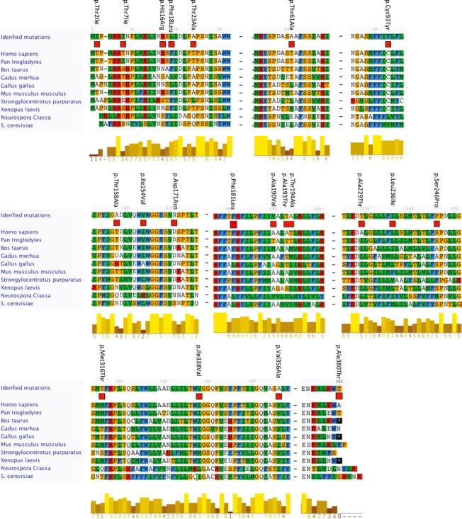
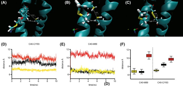
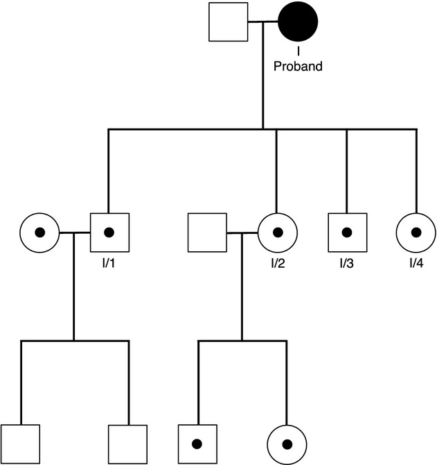
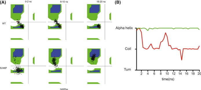
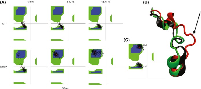
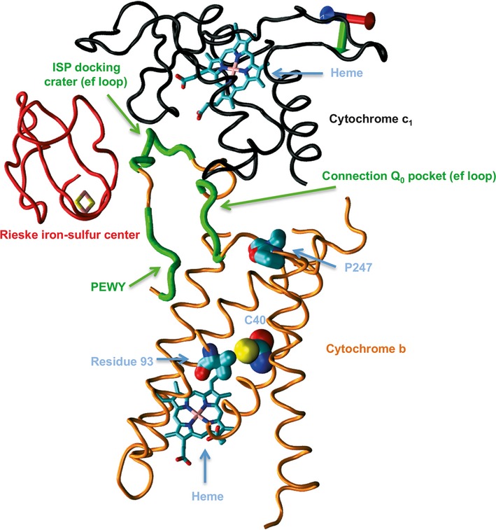
Mitochondrial dysfunction is a characteristic of heart failure. Mutations in mitochondrial DNA, particularly in MT-CYB coding for cytochrome B in complex III (CIII), have been associated with isolated hypertrophic cardiomyopathy (HCM). We hypothesized that MT-CYB mutations might play an important causal or modifying role in HCM. The MT-CYB gene was sequenced from DNA isolated from blood from 91 Danish HCM probands. Nonsynonymous variants were analyzed by bioinformatics, molecular modeling and simulation. Two germline-inherited, putative disease-causing, nonsynonymous variants: m.15024G>A; p.C93Y and m.15482T>C; p.S246P were identified. Modeling showed that the p.C93Y mutation leads to disruption of the tertiary structure of Cytb by helix displacement, interfering with protein-heme interaction. The p.S246P mutation induces a diproline structure, which alters local secondary structure and induces a kink in the protein backbone, interfering with macromolecular interactions. These molecular effects are compatible with a leaky phenotype, that is, limited but progressive mitochondrial dysfunction. In conclusion, we find that rare, putative leaky mtDNA variants in MT-CYB can be identified in a cohort of HCM patients. We propose that further patients with HCM should be examined for mutations in MT-CYB in order to clarify the role of these variants.

Keywords: Cardiomyopathy; DNA sequencing; genetic disorders; hypertrophy; mitochondria.


Figures

Figure 1
Figure 1
Cytb conservation index. Multiple alignments of the Cytb amino acid sequences with the identified nonsynonymous mutations marked. The position of the mutation is highlighted and the amino acids are numbered according to the Homo sapiens sequence. The conservation scores are generated by Jalview and reflect the conservation of the physicochemical properties of each column in the alignment.
Figure 2
Figure 2
Pedigree of family B with the p.C93Y mutation. Proband B is marked with an arrow. Individuals colored black are carriers of the MHC-β p.R1712W mutation diagnosed with HCM. Individuals with a black dot are asymptomatic MHC-β, R1712W carriers.
Figure 3
Figure 3
Molecular simulation of Cytb. The p.Y93 mutant is shown in red, p.C93 is shown in black, and p.C93 with a disulfide bridge to p.C40 is shown in yellow. Visualization of coordinates from the end of the simulations were generated using VMD and are shown for p.Y93 (A), p. C93 (B), and p.C93 with a disulfide bridge to p.C40 (C). The distances between p.C40 and p.C/Y93 (D) and between p.C40 and p.M89 (E) are shown as a function of simulation time. These values are shown combined (F). Whiskers are 2.5–97.5 percentile. Initial transients are discarded from p.C40-p.C/Y93 values, and data shown is from 7 ns and forward.
Figure 4
Figure 4
Pedigree of family I with the p.S246P mutation. Individuals with black dots are carriers of the mutation. Individuals colored black are diagnosed with HCM.
Figure 5
Figure 5
p.F245 simulated participation in secondary structure. (A) Ramachandran plots of p.F245 over the time-course of the simulation. (B) Secondary structure calculated using STRIDE during the course of the simulation is shown. Each data point on the graph corresponds to a measure of the prevalence of different secondary structures during 0.5 ns of simulation time.
Figure 6
Figure 6
Simulated structural effects of p.S246P. (A) Ramachandran plots of p.D248 over the time-course of the simulation. (B) Final structure of p.P246 (red) compared with p.S246 (green) after 20 ns. The initial backbone of both p.P246 and p.S246 is also shown (black). The black arrow denotes the kink induced by p.P246 (C). Ramachandran plot of p.P246 and p.P247 from 18–20 ns, showing the final angle configuration, compatible with a PIIR conformation.
Figure 7
Figure 7
Relevant parts of yeast Cytochrome bc1 (PDB ID: 3cxh). Cytochrome b is shown in orange. Cytochrome c1 is shown in black. The Rieske subunit is shown in red. The location of the PEWY motif, ISP docking crater, and Q0 connection on Cytochrome b is shown in green. The location of the conserved p.P247 and the location of p.C40 and residue number 93 (valine in yeast) are shown by blue arrows. Heme groups are also marked by blue arrows.

Similar articles

Cited by

References

    1. Andersen PS, Havndrup O, Hougs L, Sorensen KM, Jensen M, Larsen LA, et al. Diagnostic yield, interpretation, and clinical utility of mutation screening of sarcomere encoding genes in Danish hypertrophic cardiomyopathy patients and relatives. Hum. Mutat. 2009;30:363–370. - PubMed
    1. Andreu AL, Checcarelli N, Iwata S, Shanske S, DiMauro S. A missense mutation in the mitochondrial cytochrome b gene in a revisited case with histiocytoid cardiomyopathy. Pediatr. Res. 2000;48:311–314. - PubMed
    1. Arbustini E, Fasani R, Morbini P, Diegoli M, Grasso M, Dal Bello B, et al. Coexistence of mitochondrial DNA and beta myosin heavy chain mutations in hypertrophic cardiomyopathy with late congestive heart failure. Heart. 1998;80:548–558. - PMC - PubMed
    1. Ballana E, Govea N, Garcia R, de Cid C, Arribas C, Rosell J, et al. Detection of unrecognized low-level mtDNA heteroplasmy may explain the variable phenotypic expressivity of apparently homoplasmic mtDNA mutations. Hum. Mutat. 2008;29:248–257. - PubMed
    1. Behar DM, Villems R, Soodyall H, Blue-Smith J, Pereira L, Metspalu E, et al. The dawn of human matrilineal diversity. Am. J. Hum. Genet. 2008;82:1130–1140. - PMC - PubMed

LinkOut - more resources