. 2013 Nov;1(4):206-22.
doi: 10.1002/mgg3.24. Epub 2013 Jul 2.

Screening of CACNA1A and ATP1A2 genes in hemiplegic migraine: clinical, genetic, and functional studies

Affiliations

Screening of CACNA1A and ATP1A2 genes in hemiplegic migraine: clinical, genetic, and functional studies

Oriel Carreño et al. Mol Genet Genomic Med. 2013 Nov.

Abstract

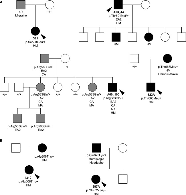
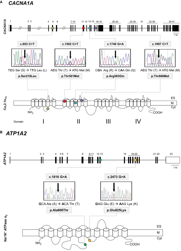
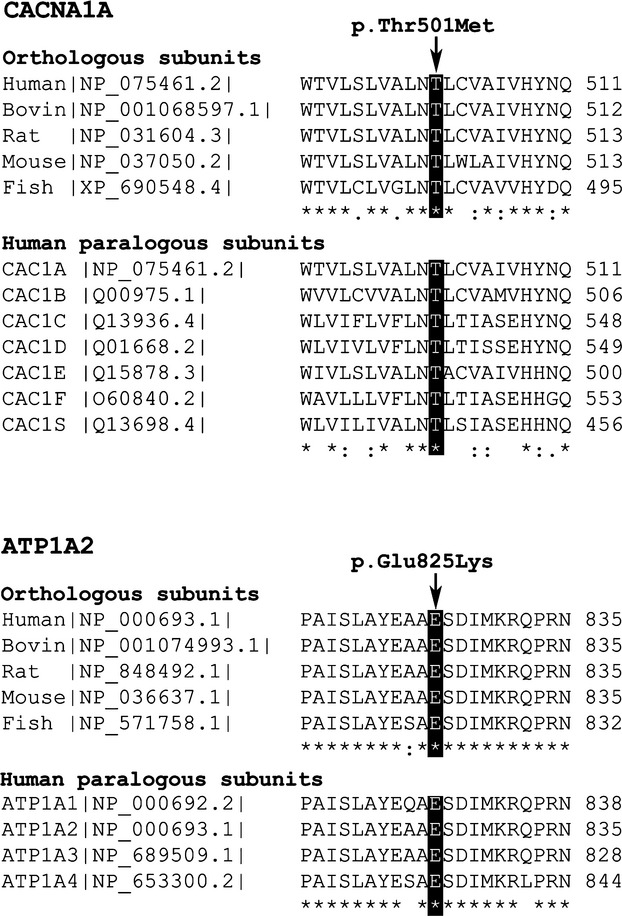
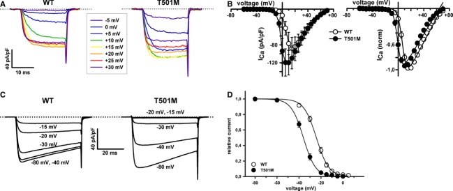
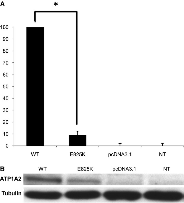
Hemiplegic migraine (HM) is a rare and severe subtype of autosomal dominant migraine, characterized by a complex aura including some degree of motor weakness. Mutations in four genes (CACNA1A, ATP1A2, SCN1A and PRRT2) have been detected in familial and in sporadic cases. This genetically and clinically heterogeneous disorder is often accompanied by permanent ataxia, epileptic seizures, mental retardation, and chronic progressive cerebellar atrophy. Here we report a mutation screening in the CACNA1A and ATP1A2 genes in 18 patients with HM. Furthermore, intragenic copy number variant (CNV) analysis was performed in CACNA1A using quantitative approaches. We identified four previously described missense CACNA1A mutations (p.Ser218Leu, p.Thr501Met, p.Arg583Gln, and p.Thr666Met) and two missense changes in the ATP1A2 gene, the previously described p.Ala606Thr and the novel variant p.Glu825Lys. No structural variants were found. This genetic screening allowed the identification of more than 30% of the disease alleles, all present in a heterozygous state. Functional consequences of the CACNA1A-p.Thr501Met mutation, previously described only in association with episodic ataxia, and ATP1A2-p.Glu825Lys, were investigated by means of electrophysiological studies, cell viability assays or Western blot analysis. Our data suggest that both these variants are disease-causing.

Keywords: ATP1A2; CACNA1A; functional studies; hemiplegic migraine; mutation analysis.


Figures

Figure 1
Figure 1
Pedigrees of patients with the identified gene variants. (A) Mutations in the CACNA1A gene. (B) Mutations in the ATP1A2 gene. Affected individuals are denoted by solid symbols; hemiplegic migraine (HM) in black and other phenotypes in gray; squares indicate males and circles females. Probands are indicated by a black arrow. Clinical characteristics are indicated below each individual (HM, migraine with hemiplegic aura; MA, migraine with aura; CA, cerebellar atrophy; EA2, episodic ataxia type 2). Gene variant carrier status is indicated below each patient when known. Mutation p.Ser218Leu appeared de novo in the affected sib.
Figure 2
Figure 2
Gene structure of CACNA1A and ATP1A2 and detection of mutations. (A) 1: CACNA1A gene structure, with black boxes indicating exons. The identified mutations causing HM are indicated by colored dots: p.Ser218Leu (light green), p.Thr501Met (red), p.Arg583Gln (blue), and p.Thr666Met (purple). 2: Protein structure and location of the identified mutations. (B) 1: ATP1A2 gene structure, with black boxes indicating exons. The newly identified mutation p.Glu825Lys causing HM is indicated by a yellow dot and p.Ala606Thr with a green dot. 2: Protein structure and location of the identified mutations. Detection of the mutations by direct sequencing of PCR products: electropherograms. Cyt, cytoplasm; M, cytoplasmic membrane; ES, extracellular space.
Figure 3
Figure 3
Protein alignment performed with ClustalW (http://www.ebi.ac.uk/Tools/msa/clustalw2) (Chenna et al. 2003). On top, the CACNA1A Thr501 residue is conserved in all the human calcium channel α1 subunits studied (CACNA1A, B, C, D, E, F, and S) and in the orthologous CACNA1A proteins of several organisms. Bottom, the ATP1A2 Glu825 residue is conserved in the four human ATP1 paralogous subunits (ATP1A1, A2, A3, and A4) and in orthologous ATP1A2 proteins of several organisms. Key: “*” identical residues; “:” conserved substitutions (same amino acid group); “.” semi-conserved substitution (similar shapes). Human: Homo sapiens; Bovin: Bos taurus; Rat: Rattus norvegicus; Mouse: Mus musculus; Fish (Zebrafish): Danio rerio.
Figure 4
Figure 4
Mutation p.Thr501Met affects activation and inactivation properties of heterologously expressed P/Q channels. (A) Current traces illustrating voltage dependence of WT (left) and p.Thr501Met (right) P/Q channels, in response to 20 msec voltage pulses. Dotted lines in the current traces indicate the zero current level. (B) Current density–voltage relationships (left panels) and normalized I–V curves (right panels) for WT (○) and p.Thr501Met (•) P/Q channels expressed in HEK 293 cells. V1/2, act and kact values were (in mV): WT (○, n = 9) 7.1 ± 0.8 and 3.3 ± 0.3; p.Thr501Met (•, n = 14) −0.04 ± 0.99 and 2.8 ± 0.3, respectively. No significant difference was found for kact values (P = 0.29). (C and D) Steady-state inactivation of WT or p.Thr501Met P/Q channels. Amplitudes of currents elicited by test pulses to +20 mV were normalized to the current obtained after a 30-sec prepulse to −80 mV and fitted by a single Boltzmann function (see Materials and Methods, eq. 2). V1/2, inact and kinact values were (in mV): WT (○, n = 10) −24.2 ± 0.9 and −5.5 ± 0.4; p.Thr501Met (•, n = 15) −35.9 ± 1 and −5 ± 0.2, respectively. No significant difference was found for kinact values (P = 0.53).
Figure 5
Figure 5
(A) Viability of HeLa cells transfected with the mutant ATP1A2 cDNA (E825K) normalized to the viability of cells transfected with the ouabain-resistant wild-type ATP1A2 cDNA (WT). NT, untransfected cells; pcDNA3.1, cells transfected with the empty vector. Four independent experiments were performed, each with triplicate measurements. The * symbol indicates the existence of significant differences between the p.Glu825Lys and the WT ATP1A2 constructs (P < 0.0003). (B) Western blot assay of the different protein extracts using anti-myc and anti-tubulin antibodies. The molecular weight of the Na+/K+-ATPase α2 subunit and tubulin are indicated. The constructs with the ATP1A2 ouabain-resistant cDNA carry the myc tag. The clone with the mutation displayed diminished band intensity.

References

    1. Adams PJ, Rungta RL, Garcia E, MacVicar AM, van den Maagdenberg BA, Snutch TP. Contribution of calcium-dependent facilitation to synaptic plasticity revealed by migraine mutations in the P/Q-type calcium channel. Proc. Natl. Acad. Sci. USA. 2010;107:18694–18699. - PMC - PubMed
    1. Alonso I, Barros J, Tuna A, Coelho J, Sequeiros J, Silveira I, et al. Phenotypes of spinocerebellar ataxia type 6 and familial hemiplegic migraine caused by a unique CACNA1A missense mutation in patients from a large family. Arch. Neurol. 2003;60:610–614. - PubMed
    1. Bassi MT, Bresolin N, Tonelli A, Nazos K, Crippa F, Baschirotto C, et al. A novel mutation in the ATP1A2 gene causes alternating hemiplegia of childhood. J. Med. Genet. 2004;41:621–628. - PMC - PubMed
    1. Battistini S, Stenirri S, Piatti M, Gelfi C, Righetti PG, Rocchi R, et al. A new CACNA1A gene mutation in acetazolamide-responsive familial hemiplegic migraine and ataxia. Neurology. 1999;53:38–43. - PubMed
    1. Bolay H, Reuter U, Dunn AK, Huang Z, Boas DA, Moskowitz MA. Intrinsic brain activity triggers trigeminal meningeal afferents in a migraine model. Nat. Med. 2002;8:136–142. - PubMed