Review
. 2014:10:679-708.
doi: 10.1146/annurev-clinpsy-032813-153716. Epub 2014 Jan 31.

The role of sleep in emotional brain function

Affiliations
Review

The role of sleep in emotional brain function

Andrea N Goldstein et al. Annu Rev Clin Psychol. 2014.

Abstract

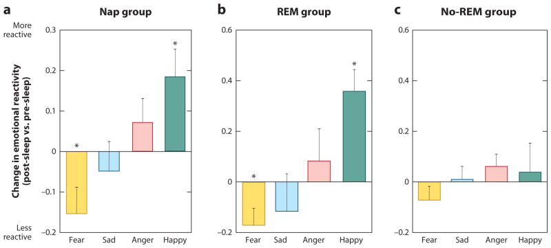
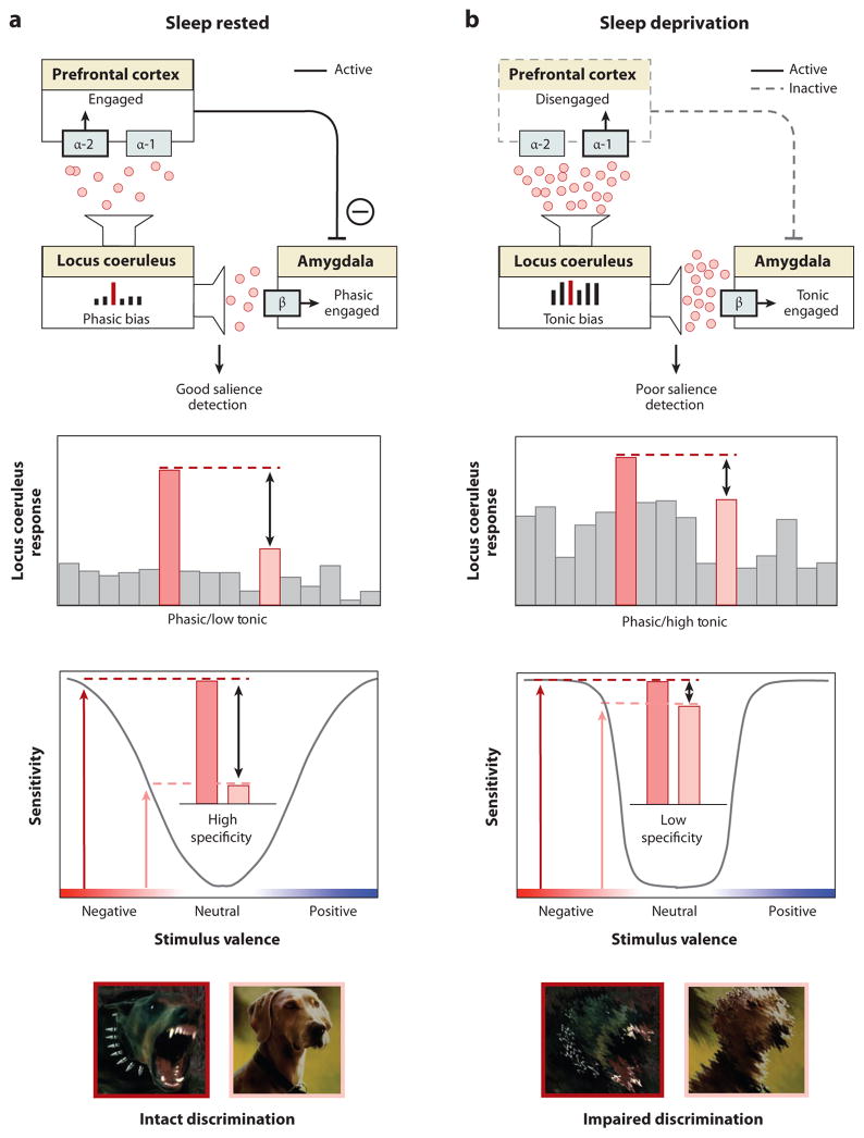
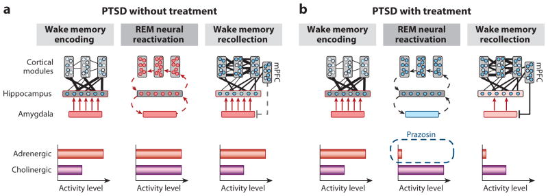
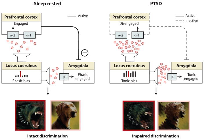
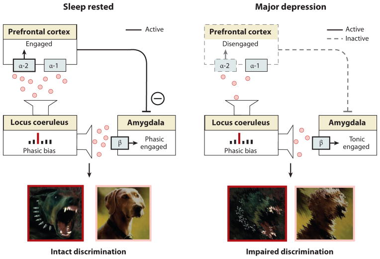
Rapidly emerging evidence continues to describe an intimate and causal relationship between sleep and emotional brain function. These findings are mirrored by long-standing clinical observations demonstrating that nearly all mood and anxiety disorders co-occur with one or more sleep abnormalities. This review aims to (a) provide a synthesis of recent findings describing the emotional brain and behavioral benefits triggered by sleep, and conversely, the detrimental impairments following a lack of sleep; (b) outline a proposed framework in which sleep, and specifically rapid-eye movement (REM) sleep, supports a process of affective brain homeostasis, optimally preparing the organism for next-day social and emotional functioning; and (c) describe how this hypothesized framework can explain the prevalent relationships between sleep and psychiatric disorders, with a particular focus on posttraumatic stress disorder and major depression.


Figures

Figure 1
Figure 1. The impact of sleep deprivation on emotional brain reactivity and functional connectivity
(a) Amygdala response to increasingly negative emotional stimuli in the Sleep deprivation and Sleep rested (control) conditions, and (b) Corresponding differences in intensity and volumetric extent of amygdala activation between the two groups (average ± s.e.m. of left and right amygdala), (c) Changes in functional connectivity between the medial prefrontal cortex (mPFC) and the amygdala. With sleep, the prefrontal lobe was significantly connected to the amygdala, regulating and exerting and inhibitory top-down control, (d) Without sleep, however, amygdala-mPFC connectivity was decreased, potentially negating top-down control and resulting in an overactive amygdala. *p < 0.01; error bars indicate s.e.m. Modified from (Yoo et al 2007).
Figure 2
Figure 2. REM sleep enhancement of negative emotional memories
(a) Offline benefit (change in memory recall for 4 hr versus 15 min old memories) across the day (wake, grey bar) or following a 90 min nap (sleep, filled bar); (b) Correlation between the amount of offline emotional memory improvement in the nap group (i.e. the offline benefit expressed in filled bar of figure a), and the amount of REM sleep obtained within the nap; (c) Correlation (Pearson’s r-value) between offline benefit for emotional memory in the sleep group (expressed in filled bar of figure a) and the relative right versus left prefrontal spectral-band power ([electrode F4 – electrode F3]) within the delta, alpha, theta and beta spectral bands, expressed in average 0.5 Hz bin increments. Correlation strength is represented by the color range, demonstrating significant correlations within the theta frequency band (hot colors), and (d) exhibiting a maximum significance at the 5.75 Hz bin. *p < 0.05; error bars indicate s.e.m. Modified from (Nishida et al 2009).
Figure 3
Figure 3. The sleep to forget and sleep to remember (SFSR) model;
(a) Neural dynamics. Emotional memory formation involves the encoding of hippocampal-bound cortical information, facilitated by the amygdala and high concentrations of aminergic activity. During REM sleep, these neural structures are reactivated, supporting the reprocessing of emotional memories. However, this occurs in a brain-state with dramatically reduced adrenergic activity, allowing for both cortical strengthening (consolidation), dissipation of previously associated emotion (visceral tone), and reestablished mPFC-amygdala regulatory control. Cross-connectivity between structures is represented by number and thickness of lines. Circles within cortical and hippocampal structures represent information nodes; shade strength reflects extent of connectivity. Fill of amygdala and arrow thickness represents influence upon the hippocampus. (b) Conceptual outcome. Through multiple iterations of this REM-mechanism across one or multiple nights, such sleep-dependent reprocessing results in long-term strengthening of salient memories, yet a dissipation of the emotional charge. Thus, sleep transforms an emotional memory into a memory of an emotional event, that itself is no longer emotional.
Figure 4
Figure 4. REM sleep depotentiates amygdala reactivity to prior emotional experiences
(a) Change in emotion reactivity: group x test session interaction in bilateral amygdala (blue), demonstrating a significant decrease in activity across a night of sleep in the sleep group, yet an increase in the wake group across a day of wake. (b) Change in functional connectivity: group x test session interaction in amygdala-ventromedial prefrontal cortex (vmPFC) connectivity (yellow), demonstrating increased connectivity from after a night of sleep yet decreased coupling after an equivalent time of wake. (c) Topographical Spearman’s correlation (ρ) plot of the relationship between electroencephalographic (EEG) gamma power during rapid-eye movement (REM) sleep and the extent of overnight emotional reactivity decrease across a night of sleep, with lower levels of prefrontal gamma activity (marked by white circles) predicting a larger overnight decrease in emotional reactivity. * p < 0.05. Modified from (van der Helm et al 2011).
Figure 5
Figure 5. Differential impact of REM sleep on emotional reactivity
Difference in mean ratings between the pre-sleep and post-sleep test sessions across 4 emotion categories (fear, sad, anger, and happy) for (a) the Nap group overall performance, (b) only for those in the Nap group who obtained REM sleep, and (c) only for those in the Nap group who did not obtain REM sleep. Within-group comparisons (symbol above individual bars) reflect paired t-test significance (relative to null) at *<0.05 and **<0.01. Error bars represent s.e.m. Modified from (Gujar et al 2010).
Figure 6
Figure 6. REM sleep emotion recalibration model
(a) Under normal sleep rested conditions, the overnight reduction of noradrenaline levels promoted by REM sleep restores noradrenergic tone, resulting in a low tonic, high phasic locus coeruleus activity. This consequently primes downstream amygdala and PFC structures that, together with the locus coeruleus, form part of an emotional salience detection network (upper panel). Moderate next-day levels of noradrenaline, restored by REM sleep, induce PFC engagement through activation of the α-2 receptor, which in turn enables top-down PFC inhibition of the amygdala, reducing the likelihood of a non-specific amygdala response. Further, low tonic activity in locus coeruleus enhances the fidelity of phasic responses to external emotional stimuli, relative to the low baseline firing, promoting sensitivity to detect truly emotional stimuli, while also retaining the ability to discriminate between degrees of emotional salience (specificity) (middle panel). The functional outcome of this REM sleep recalibrated salience network state is the ability to discriminate between, and appropriately react to, emotionally salient events (e.g., threatening, aggressive dog) from non-salient stimuli (e.g., non-threatening dog) (lower panel). b) In contrast, sleep deprivation, results in high tonic locus coeruleus firing mode and increased noradrenaline concentrations, consequently impairing salience detection. Specifically, elevated noradrenaline levels increase binding to the inhibitory α-1 receptor in the PFC (upper panel). Unlike binding to α-2 receptors, α-1 receptor binding impairs PFC functioning and so reduces top-down inhibition of the amygdala. This disinhibition results in a non-specific, generalized amygdala responding to both emotionally salient and non-salient stimuli. Such impairments are further exacerbated by high tonic baseline locus coeruleus activity, which reduces the ability to differentiate phasic signals from high baseline activity (i.e. reduced signal-to-noise). In this mode of activity, the locus coeruleus responds non-specifically to both emotionally salient and non-emotionally salient information (middle panel). Together, these alterations to the emotional salience network caused by sleep loss impair the accurate discrimination of threatening from non-threatening stimuli, resulting in persistent sensitivity and in impaired emotional specificity (lower panel). Noradrenaline alpha-1 receptor (α-1), noradrenaline alpha-2 receptor (α-2) and noradrenaline beta receptor (β).
Figure 7
Figure 7. REM Sleep to forget sleep to remember model applied to PTSD
(a) REM sleep in PTSD without treatment, characterized by a pathological persistence of central brain adrenergic activity (note difference in middle panel to middle panel of Figure 3a). Elevated adrenergic activity during REM sleep prevents the depotentiation of the emotion tone associated with the salient, including traumatic, experiences. (b) REM sleep in PTSD with treatment by Prazosin, allowing for the reduction in adrenergic levels during REM sleep (note difference in middle panel between a and b). Consequently, there is the restored opportunity for the dissipation of emotion from the prior trauma experiences, preventing hyperarousal reactions during subsequent post-sleep memory recall.
Figure 8
Figure 8. REM Sleep Emotion Recalibration model applied to PTSD
Elevated noradrenergic activity during REM sleep in PTSD prevents the next day recalibration of the associated salience network, impairing optimal emotion discrimination, similar to the state if sleep deprivation (see also sleep deprivation profile in figure 6)
Figure 9
Figure 9. REM Sleep Emotion Recalibration model applied to major depression
Exaggerated REM sleep qualities in major depression produce an excessive blunting in next-day noradrenergic activity, resulting in too little noradrenaline for appropriate emotional salience detection.

Similar articles

Cited by

References

    1. Anderson C, Platten CR. Sleep deprivation lowers inhibition and enhances impulsivity to negative stimuli. Behav Brain Res. 2011;217:463–6. - PubMed
    1. Appelhans B, Luecken L. Heart rate variability as an index of regulated emotional responding. Review of General Psychology. 2006;10:229–40.
    1. Armitage R. Sleep and circadian rhythms in mood disorders. Acta Psychiatr Scand Suppl. 2007:104–15. - PubMed
    1. Arnedt JT, Conroy DA, Brower KJ. Treatment options for sleep disturbances during alcohol recovery. J Addict Dis. 2007;26:41–54. - PMC - PubMed
    1. Aston-Jones G, Cohen JD. An integrative theory of locus coeruleus-norepinephrine function: adaptive gain and optimal performance. Annu Rev Neurosci. 2005;28:403–50. - PubMed

Publication types

MeSH terms