

Modulation of experimental atopic dermatitis by topical application of Gami-Cheongyeul-Sodok-Eum
- PMID: 24499290
- PMCID: PMC3832229
- DOI: 10.1186/1472-6882-13-312
Modulation of experimental atopic dermatitis by topical application of Gami-Cheongyeul-Sodok-Eum
Abstract
Background: Gami-Cheongyeul-Sodok-Eum (GCSE), an herbal formula of traditional Korean medicine, comprises nine herb components. GCSE has various biological activities such as anti-inflammatory, anti-bacterial and anti-viral activities. However, it is still unclear whether GCSE has any immunomodulatory effect on atopic dermatitis (AD).
Methods: GCSE was treated to primary B cells and CD4+ T cells isolated from atopic mice to compare its inhibitory effects on IgE secretion and cytokine expression. Experimental AD was established by alternative treatment of 2, 4-dinitrochlorobenzene (DNCB) and house dust mite extract to the ears of BALB/c mice. GCSE was topically applied to ears of atopic mice every day for 3 weeks. AD progression was analyzed by measuring ear thickness, serum IgE level, histological examination of ear tissue by H&E staining and cytokine profile of CD4+ T cells and CD19+ B cells by real time PCR and ELISA.
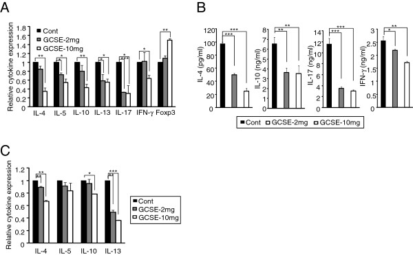
Results: Treatment of GCSE significantly reduced IgE production and expression of AD associated pathogenic cytokines such as IL-4, IL-5, IL-10, IL-13, IL-17, TNF-α, and IFN-γ by lymphocytes isolated from AD-induced mice. Topical application of GCSE on the ears of AD-induced mice significantly reduced ear thickness, clinical score and lymphocytes infiltration to ears as compared to control group. GCSE treatment also reduced serum IgE level and the levels of major pathogenic cytokines such as IL-4, IL-5, IL-10, IL-13 and IL-17. In addition, GCSE treatment significantly increased Foxp3 expression level.
Conclusions: The protective effect of GCSE in experimental AD is mediated by inhibition of IgE production, by reduction in the levels of pathogenic cytokines and by induction of Foxp3, all of which are suggesting the beneficial effect of GCSE on modulating atopic dermatitis.
Figures


Similar articles
-
Topical application of Taglisodog-eum inhibits the development of experimental atopic dermatitis.J Ethnopharmacol. 2013 Jan 30;145(2):536-46. doi: 10.1016/j.jep.2012.11.026. Epub 2012 Dec 2. J Ethnopharmacol. 2013. PMID: 23211659
-
Immunomodulatory effect of water soluble extract separated from mycelium of Phellinus linteus on experimental atopic dermatitis.BMC Complement Altern Med. 2012 Sep 18;12:159. doi: 10.1186/1472-6882-12-159. BMC Complement Altern Med. 2012. PMID: 22988890 Free PMC article.
-
Suppression of dust mite extract and 2,4-dinitrochlorobenzene-induced atopic dermatitis by the water extract of Lindera obtusiloba.J Ethnopharmacol. 2011 Sep 1;137(1):802-7. doi: 10.1016/j.jep.2011.06.043. Epub 2011 Jul 5. J Ethnopharmacol. 2011. PMID: 21762765
-
Management of Atopic Dermatitis Via Oral and Topical Administration of Herbs in Murine Model: A Systematic Review.Front Pharmacol. 2022 May 24;13:785782. doi: 10.3389/fphar.2022.785782. eCollection 2022. Front Pharmacol. 2022. PMID: 35685636 Free PMC article.
-
Current Insight into the Role of IL-35 and Its Potential Involvement in the Pathogenesis and Therapy of Atopic Dermatitis.Int J Mol Sci. 2022 Dec 11;23(24):15709. doi: 10.3390/ijms232415709. Int J Mol Sci. 2022. PMID: 36555351 Free PMC article. Review.
Cited by
-
Galectin-9 Induced by Dietary Prebiotics Regulates Immunomodulation to Reduce Atopic Dermatitis Symptoms in 1-Chloro-2,4-Dinitrobenzene (DNCB)-Treated NC/Nga Mice.J Microbiol Biotechnol. 2020 Sep 28;30(9):1343-1354. doi: 10.4014/jmb.2005.05017. J Microbiol Biotechnol. 2020. PMID: 32699202 Free PMC article.
-
Interconnection of the Gut-Skin Axis in NC/Nga Mouse with Atopic Dermatitis: Effects of the Three Types of Bifidobacterium bifidum CBT-BF3 (Probiotics, Postbiotics, and Cytosine-Phosphate-Guanine Oligodeoxynucleotide) on T Cell Differentiation and Gut Microbiota.Food Sci Anim Resour. 2024 Nov;44(6):1417-1439. doi: 10.5851/kosfa.2024.e100. Epub 2024 Nov 1. Food Sci Anim Resour. 2024. PMID: 39554831 Free PMC article.
References
Publication types
MeSH terms
Substances
LinkOut - more resources
Full Text Sources
Other Literature Sources
Research Materials
