. 2014 Feb 6:10:10.
doi: 10.1186/1744-8069-10-10.

Toll like receptor (TLR)-4 as a regulator of peripheral endogenous opioid-mediated analgesia in inflammation

Affiliations

Toll like receptor (TLR)-4 as a regulator of peripheral endogenous opioid-mediated analgesia in inflammation

Reine-Solange Sauer et al. Mol Pain. .

Abstract

Background: Leukocytes containing opioid peptides locally control inflammatory pain. In the early phase of complete Freund's adjuvant (CFA)-induced hind paw inflammation, formyl peptides (derived e.g. from Mycobacterium butyricum) trigger the release of opioid peptides from neutrophils contributing to tonic basal antinociception. In the later phase we hypothesized that toll-like-receptor-(TLR)-4 activation of monocytes/macrophages triggers opioid peptide release and thereby stimulates peripheral opioid-dependent antinociception.

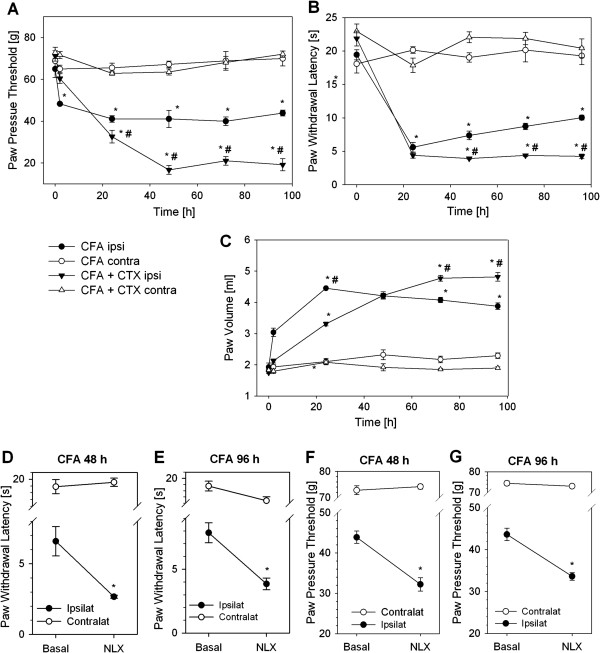
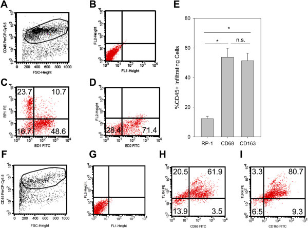
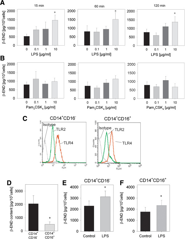
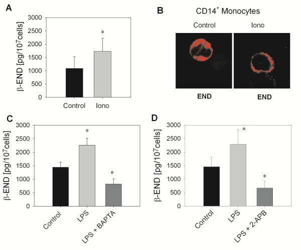
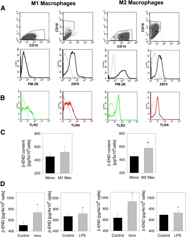
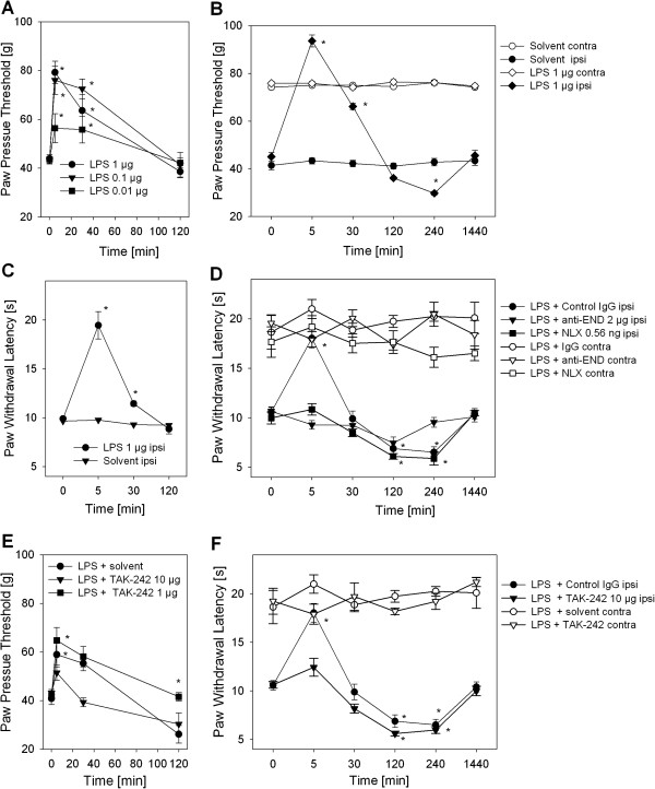
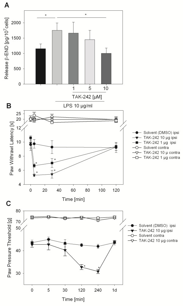
Results: In Wistar rats with CFA hind paw inflammation in the later inflammatory phase (48-96 h) systemic leukocyte depletion by cyclophosphamide (CTX) or locally injected naloxone (NLX) further decreased mechanical and thermal nociceptive thresholds. In vitro β-endorphin (β-END) content increased during human monocyte differentiation as well as in anti-inflammatory CD14+CD16- or non-classical M2 macrophages. Monocytes expressing TLR4 dose-dependently released β-END after stimulation with lipopolysaccharide (LPS) dependent on intracellular calcium. Despite TLR4 expression proinflammatory M1 and anti-inflammatory M2 macrophages only secreted opioid peptides in response to ionomycin, a calcium ionophore. Intraplantar injection of LPS as a TLR4 agonist into the inflamed paw elicited an immediate opioid- and dose-dependent antinociception, which was blocked by TAK-242, a small-molecule inhibitor of TLR4, or by peripheral applied NLX. In the later phase LPS lowered mechanical and thermal nociceptive thresholds. Furthermore, local peripheral TLR4 blockade worsened thermal and mechanical nociceptive pain thresholds in CFA inflammation.

Conclusion: Endogenous opioids from monocytes/macrophages mediate endogenous antinociception in the late phase of inflammation. Peripheral TLR4 stimulation acts as a transient counter-regulatory mechanism for inflammatory pain in vivo, and increases the release of opioid peptides from monocytes in vitro. TLR4 antagonists as new treatments for sepsis and neuropathic pain might unexpectedly transiently enhance pain by impairing peripheral opioid analgesia.


Figures

Figure 1
Figure 1
Increased hyperalgesia by leukocyte depletion or peripheral opioid receptor blockade in inflammatory pain. Male Wistar rats were leukocyte depleted with cyclophosphamide (CTX, 100 mg/kg and 50 mg/kg BW on day −3, -1 and 1). Complete Freund’s adjuvant (CFA) was injected i.pl. at day 0. Paw pressure thresholds (A), paw withdrawal latency (B) and paw volume (C) were measured at 0, 24, 48, 72 and 96 h after induction of inflammation in the ipsilateral (ipsi) and contralateral (contra) paw (no CTX n = 3–6, CTX n = 6–8,. * p < 0.05 vs. 0 h, # p < 0.05 vs. CFA ipsilateral, Two Way RM ANOVA, Student Newman Keuls). CTX alone did not change paw pressure thresholds [28]. (D, F) After 48 h and (E, G) 96 h of CFA hind-paw inflammation the opioid receptor antagonist naloxone (NLX, 0.56 ng) was injected i.pl. Thermal (D, E) and mechanical (F, G) nociceptive thresholds were determined before and 10 min after treatment and compared to contralateral side (n = 4–6, * p < 0.05, * vs. basal, Two Way ANOVA, Student-Newman-Keuls). No change was seen in saline injected rats before [14]. All data are presented as MEAN ± SEM.
Figure 2
Figure 2
Characterization of infiltrating leukocytes in the inflamed paw. Rats were injected with CFA for 4 d. (A) Cells suspensions from subcutaneous paw tissue were first stained for CD45 to gate on hematopoetic cells (x-axis: forward scatter [size], y-axis: CD45-PerCp-Cy5). (B) The dot plot (x-axis: FITC, y-axis: PE) shows unstained controls from cells gated on CD45. CD45+ cells were stained for RP-1 (neutrophils), CD68 (M1, proinflammatory macrophages) (C, x-axis: CD68-FITC, y-axis: RP-1-PE) as well as and CD163 (M2, antiinflammatory macrophages) (D, x-axis: CD163-FITC, y-axis: PE). Representative examples are shown (n = 8). (E) The percentage of infiltrating leukocyte subpopulations were measured (n = 8, ANOVA on Ranks, Student-Newman-Keuls). (F-I) TLR4-Expression in the paw was analyzed in leukocytes gated on CD45+ (F, x-axis: forward scatter, y-axis: CD45-PerCp-Cy5 fluorescence). TLR4-Expression in CD68+ M1 macrophages (H, x-axis: CD68-FITC, y-axis: TLR4-PE) and in CD163+ M2 macrophages (I, x-axis: CD163-FITC, y-axis: TLR4-PE) and compared to the unstained control (G, unstained control, x-axis: FITC fluorescence, y-axis: PE fluorescence). Representative examples are shown (n = 2).
Figure 3
Figure 3
Release of opioid peptides from monocytes in response to TLR4 but not TLR2 stimulation. (A, B) Monocytes were isolated from human buffy coats and purified for CD14. Release of β-endorphin (β-END) was quantified after incubation for 15 or 120 min with different doses of LPS or Pam3CSK4 (n = 7–9, * p < 0.05, One way RM ANOVA, Student-Newman-Keuls). (C) Monocytes were separated into CD14+CD16- and CD14+CD16+ monocytes. Expression of TLR2 (green line) and TLR4 (red line) was analyzed using flow cytometry in both populations. Isotype controls are shown (black line) (representative example, n = 3). (D) β-END content after lysis of unstimulated cells was measured by ELISA in these populations, CD14+CD16- (black) and CD14+CD16+ (black striped) monocytes (n = 8, * p < 0.05 paired t-test) (E,F) CD14+CD16- and CD14+CD16+ monocytes were stimulated for 15 min with 10 μg/ml LPS (red) compared to stimulation with solvent medium (black). β-END was analyzed in the supernatant (n = 8, * p < 0.05, paired t-test). All data are presented as MEAN ± SEM.
Figure 4
Figure 4
Calcium requirement of TLR4-stimulated β-END release. (A) Release of β-END in the supernatant was quantified after incubation of CD14+ monocytes for 15 min with 10 μM ionomycin (iono) or medium (control) (n = 3, * p < 0.05, paired t-test). (B) CD14+ monocytes were stimulated as described before and stained on cytospins using a ββ-END antibody (red) and confocal analysis (representative example, n = 3). (C-E) CD14+ monocytes were preincubated with BAPTA-AM (100 μM, n = 24–28, * p < 0.05, One Way RM ANOVA, Student-Newman-Keuls, C), 2-APB (100 nM, n = 11 * p < 0.05, One Way RM ANOVA, Student-Newman-Keuls, D) for 10 min before the addition of 10 μg/ml LPS for 15 min. Secretion of β-END was quantified in the supernatant. All data are presented as MEAN ± SEM.
Figure 5
Figure 5
Content and release of β-END in macrophage differentiation. CD14+ monocytes were in vitro differentiated into M1 macrophages with GM-CSF (left column) and M2 macrophages with M-CSF and IL-10 (right column). Surface marker expression of CD16, CD14 as well as PM-2 K and 25 F9 as well as TLR2 and TLR4 were evaluated by flow cytometry. (A) Expression of CD16, CD14 as well as PM-2 K and 25 F9 on M1 and M2 macrophages as well as (B) TLR4 (red line) and TLR2 (green line). Representative examples were shown; isotype controls are depicted in grey lines (representative examples). (C) CD14+ monocytes (Mono) before differentiation and M1 and M2 macrophages (Mac) from the same individual after 6–7 d of culture were lysed and intracellular β-END content measured by ELISA (n = 14, * p < 0.05, paired t-test). (D) Release of β-END after incubation with 10 μM ionomycin (iono) or 10 μg/ml lipopolysaccharide (LPS) for 15 min from M1 and M2 macrophages was quantified in the supernatant (n = 21–24 for LPS and n = 7–8 for ionomycin, * p < 0.05, paired t-test). All data are presented as MEAN ± SEM.
Figure 6
Figure 6
Opioid-dependent LPS-induced antinociception in inflammatory pain. All rats were injected with CFA i.pl. 4 d before further treatment. (A) Mechanical nociceptive thresholds were measured before and 5–120 min after injection of 100 μl LPS (0.01-1 μg) i.pl. (n = 6, * vs. 0 min, p < 0.05, Two way RM ANOVA, Student-Newman-Keuls). (B, C) Rats were treated with 1 μg LPS i.pl. and thermal nociceptive as well as mechanical thresholds were determined after indicated time points (n = 6, * vs. 0 min, * p < 0.05, Two way RM ANOVA, Student-Newman-Keuls). (D) Thermal nociceptive thresholds were measured after 5 min – 1 d in rats after 1 μg LPS i.pl. and as well as NLX (0.56 ng) or anti-β-endorphin antibody (anti-END, 2 μg) compared to control antibody (IgG2 μg, all black symbols). Contralateral paws from rats in each treatment group are shown for comparison (white symbols). (E,F) The effect of the small molecule TLR4 inhibitor, TAK-242 i.pl. (1, 10 μg/ml) on LPS-induced antinociception was evaluated in mechanical and thermal nociceptive thresholds. (E) Paw pressure thresholds in inflamed paws (ipsi, black symbols) were obtained after 5–120 min (n = 3–7). (F) Thermal nociceptive thresholds were measured in inflamed paws (ipsi, black symbols) rats after 1 μg LPS and 10 μg TAK-242 i.pl. at indicated time points compared to solvent (n = 6, * vs. 0 min, p < 0.05, Two way RM ANOVA, Student-Newman-Keuls). Contralateral (contra) paws are shown seen for comparison (white symbols). The data from the treatment group LPS + control IgG (black circles) is displayed for comparison from graph 6D. All data are presented as MEAN ± SEM. always n = 6 except when indicated otherwise, * vs. 0 min, p < 0.05, Two way RM ANOVA, Student-Newman-Keuls).
Figure 7
Figure 7
Hyperalgesia by TLR4 blockade in inflammatory pain. (A) Purified undifferentiated CD14+ human monocytes were incubated with 10 μg/ml LPS and a TLR4 inhibitor (TAK-242) in different doses. β-END release was quantified in the supernatant after 15 min (n = 6, * vs. LPS, *p <0.05, One Way RM ANOVA, Student-Newman-Keuls). (B, C) Rats with 4 d CFA inflammation were treated with TAK-242 i.pl. (1 μg, black triangle, and 10 μg, black square) and paw withdrawal latency (B) or paw pressure thresholds (C) were measured subsequently at indicated time points and compared to solvent (5% DMSO, black circle) in the inflamed paws (ipsilateral, ipsi). Paw pressure thresholds and paw withdrawal latencies from contralateral (contra, white symbols) paws are shown for comparison (n = 3–6, * vs. 0 min, * p < 0.05, Two way RM ANOVA, Student-Newman-Keuls). All data are presented as MEAN ± SEM.

Similar articles

Cited by

References

    1. Djouhri L, Koutsikou S, Fang X, McMullan S, Lawson SN. Spontaneous pain, both neuropathic and inflammatory, is related to frequency of spontaneous firing in intact C-fiber nociceptors. J Neurosci. 2006;26:1281–1292. doi: 10.1523/JNEUROSCI.3388-05.2006. - DOI - PMC - PubMed
    1. Rittner HL, Machelska H, Stein C. Leukocytes in the regulation of pain and analgesia. J Leukoc Biol. 2005;79:1215–1222. - PubMed
    1. Liu T, Gao YJ, Ji RR. Emerging role of Toll-like receptors in the control of pain and itch. Neurosci Bull. 2012;28:131–144. doi: 10.1007/s12264-012-1219-5. - DOI - PMC - PubMed
    1. Qi J, Buzas K, Fan H, Cohen JI, Wang K, Mont E, Klinman D, Oppenheim JJ, Howard OM. Painful pathways induced by TLR stimulation of dorsal root ganglion neurons. J Immunol. 2011;186:6417–6426. doi: 10.4049/jimmunol.1001241. - DOI - PMC - PubMed
    1. Tedesco LS, Fuseler J, Grisham M, Wolf R, Roerig SC. Therapeutic administration of nitric oxide synthase inhibitors reverses hyperalgesia but not inflammation in a rat model of polyarthritis. Pain. 2002;95:215–223. doi: 10.1016/S0304-3959(01)00402-X. - DOI - PubMed

Publication types

MeSH terms

LinkOut - more resources