

Redescription of Onchocerca lupi (Spirurida: Onchocercidae) with histopathological observations
- PMID: 24499611
- PMCID: PMC3818983
- DOI: 10.1186/1756-3305-6-309
Redescription of Onchocerca lupi (Spirurida: Onchocercidae) with histopathological observations
Abstract
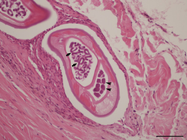
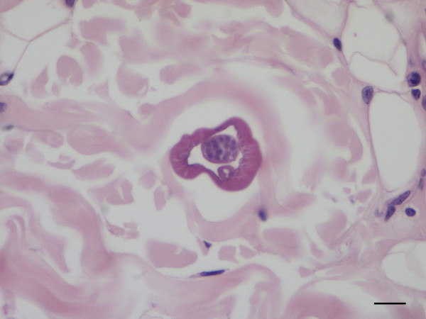
Background: Onchocerca lupi is a dog parasite of increasing zoonotic concern, with new human cases diagnosed in Turkey, Tunisia, Iran, and the United States. Information about the morphology of this nematode is scant and a detailed re-description of this species is overdue. In addition, histopathological data of potential usefulness for the identification of O. lupi infections are provided.
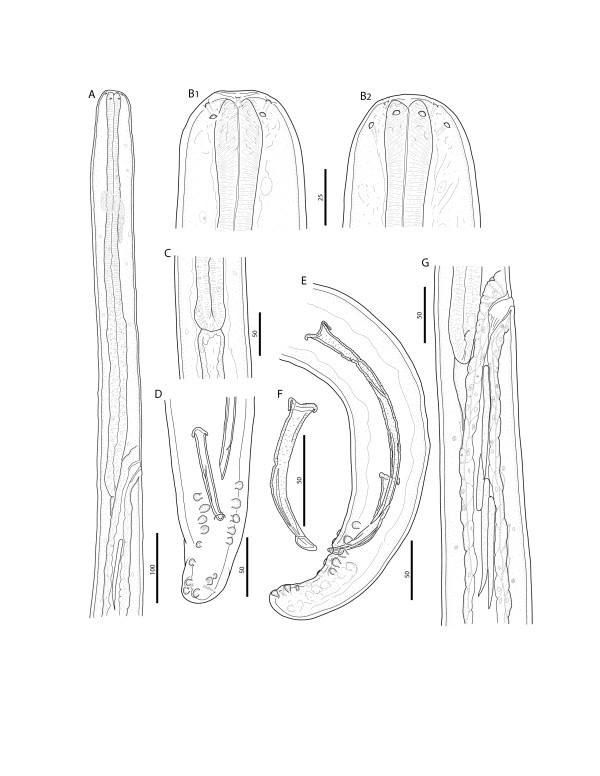
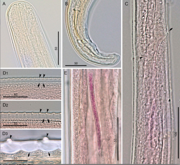
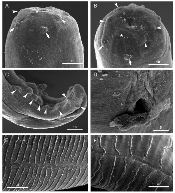
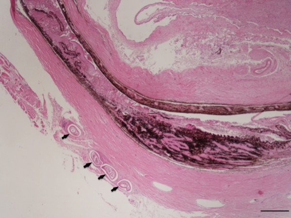
Methods: Male and female nematodes, collected from the connective tissue of a dog, were examined using light microscopy and scanning electron microscopy (SEM), and an histological evaluation was performed on biopsy samples from periocular tissues.
Results: The morphological identification was confirmed by molecular amplification and partial sequencing of cytochrome oxidase subunit 1 gene. This study provides the first comprehensive morphological and morphometric description of O. lupi from a dog based on light microscopy, SEM, molecular characterization, and histological observations.
Conclusions: Data herein presented contribute to a better understanding of this little known parasitic zoonosis, whose impact on human and animal health is still underestimated. The presence of granulomatous reactions only around the female adult suggests that the release of microfilariae from the uterus might be the cause of the inflammatory reaction observed.
Figures

Similar articles
-
Cutaneous distribution and circadian rhythm of Onchocerca lupi microfilariae in dogs.PLoS Negl Trop Dis. 2013 Dec 12;7(12):e2585. doi: 10.1371/journal.pntd.0002585. eCollection 2013. PLoS Negl Trop Dis. 2013. PMID: 24349594 Free PMC article.
-
A real-time PCR tool for the surveillance of zoonotic Onchocerca lupi in dogs, cats and potential vectors.PLoS Negl Trop Dis. 2018 Apr 4;12(4):e0006402. doi: 10.1371/journal.pntd.0006402. eCollection 2018 Apr. PLoS Negl Trop Dis. 2018. PMID: 29617361 Free PMC article.
-
Aberrant laryngeal location of Onchocerca lupi in a dog.Parasitol Int. 2016 Jun;65(3):218-20. doi: 10.1016/j.parint.2015.12.010. Epub 2015 Dec 28. Parasitol Int. 2016. PMID: 26732654
-
Clinical case presentation and a review of the literature of canine onchocercosis by Onchocerca lupi in the United States.Parasit Vectors. 2015 Feb 8;8:89. doi: 10.1186/s13071-015-0699-3. Parasit Vectors. 2015. PMID: 25884672 Free PMC article. Review.
-
Onchocercosis: a newly recognized disease in dogs.Vet Parasitol. 2008 Jan 25;151(1):1-13. doi: 10.1016/j.vetpar.2007.09.008. Epub 2007 Oct 24. Vet Parasitol. 2008. PMID: 17951007 Review.
Cited by
-
Confirmed cases of human Onchocerca lupi infection: a systematic review of an emerging threat.Parasitol Res. 2021 Nov;120(11):3633-3644. doi: 10.1007/s00436-021-07309-2. Epub 2021 Sep 14. Parasitol Res. 2021. PMID: 34519871
-
Cutaneous distribution and circadian rhythm of Onchocerca lupi microfilariae in dogs.PLoS Negl Trop Dis. 2013 Dec 12;7(12):e2585. doi: 10.1371/journal.pntd.0002585. eCollection 2013. PLoS Negl Trop Dis. 2013. PMID: 24349594 Free PMC article.
-
Diversity of Cercopithifilaria species in dogs from Portugal.Parasit Vectors. 2014 Jun 5;7:261. doi: 10.1186/1756-3305-7-261. Parasit Vectors. 2014. PMID: 24898125 Free PMC article.
-
Zoonotic Onchocerca lupi infection in dogs, Greece and Portugal, 2011-2012.Emerg Infect Dis. 2013 Dec;19(12):2000-3. doi: 10.3201/eid1912.130264. Emerg Infect Dis. 2013. PMID: 24274145 Free PMC article.
-
Onchocerciasis caused by Onchocerca lupi: an emerging zoonotic infection. Systematic review.Parasitol Res. 2015 Jul;114(7):2401-13. doi: 10.1007/s00436-015-4535-7. Epub 2015 May 20. Parasitol Res. 2015. PMID: 25990062
References
Publication types
MeSH terms
Substances
Associated data
- Actions
- Actions
LinkOut - more resources
Full Text Sources
Other Literature Sources
