. 2014 Mar 13;507(7491):190-4.
doi: 10.1038/nature12930. Epub 2014 Feb 5.

Alveolar progenitor and stem cells in lung development, renewal and cancer

Affiliations

Alveolar progenitor and stem cells in lung development, renewal and cancer

Tushar J Desai et al. Nature. .

Abstract

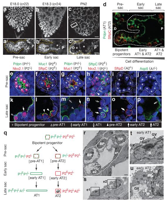
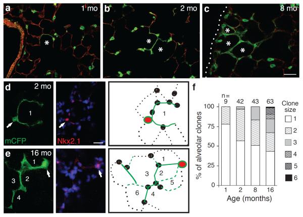
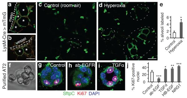
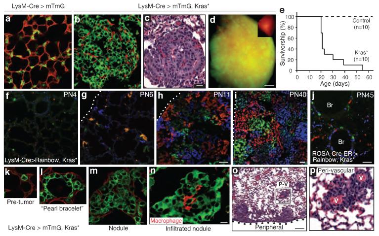
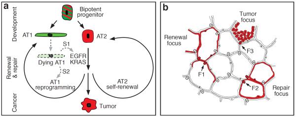
Alveoli are gas-exchange sacs lined by squamous alveolar type (AT) 1 cells and cuboidal, surfactant-secreting AT2 cells. Classical studies suggested that AT1 arise from AT2 cells, but recent studies propose other sources. Here we use molecular markers, lineage tracing and clonal analysis to map alveolar progenitors throughout the mouse lifespan. We show that, during development, AT1 and AT2 cells arise directly from a bipotent progenitor, whereas after birth new AT1 cells derive from rare, self-renewing, long-lived, mature AT2 cells that produce slowly expanding clonal foci of alveolar renewal. This stem-cell function is broadly activated by AT1 injury, and AT2 self-renewal is selectively induced by EGFR (epidermal growth factor receptor) ligands in vitro and oncogenic Kras(G12D) in vivo, efficiently generating multifocal, clonal adenomas. Thus, there is a switch after birth, when AT2 cells function as stem cells that contribute to alveolar renewal, repair and cancer. We propose that local signals regulate AT2 stem-cell activity: a signal transduced by EGFR-KRAS controls self-renewal and is hijacked during oncogenesis, whereas another signal controls reprogramming to AT1 fate.


Figures

Figure 1
Figure 1. Development of alveolar type 1 (AT1) and AT2 cells from bipotent progenitors
(a–c) Mouse lung lobe tips stained for E-cadherin (E, embryonic day; cr, crown-rump length in mm; PN, postnatal day). (a'–c') Sacculation (sac; asterisks) proceeds proximally (P) to distally (D) along the airway. Bars, 100um (a–c), 20um (a'–c'). (d) Staining for AT1 (Pdpn) and AT2 (SftpC) markers at E18.3 shows co-expression in pre-sacculation zone and restriction in late sacculation zone. Bar, 10um. (e–p) Tips imaged in pre-sacculation (e–j) and late sacculation (k–p) zones identifies six classes of marker expression profiles (P1E,P1L,A1L,P2E,P2L,A2L; Extended Data Table 1). Bar, 20um. (q) Inferred differentiation pathways showing changes in AT1 (green) and AT2 (red) marker classes. Oval, lamellar body. (r,s) Electron micrographs at E18.3 showing early (u) and late (v) sacculation zones. BP, bipotent progenitors; e1, early AT1 cells; e2, early AT2 cells. Bar, 2um. (t,u) Boxed regions in S showing (e1) an early AT1 (squamous, glycogen vacuoles (GV) without lamellar bodies (LB)) and (e2) an early AT2 (cuboidal, GV, LB). N, nucleus; bar, 0.5um.
Figure 2
Figure 2. Mature AT2 cells renew AT1 cells in clonal foci
(a–c) AT2 lineage-labeled AT1 cell foci (green) enlarge with aging, incorporating adjacent alveoli (asterisks). Dotted line, mesothelium. Bar, 150um (a–c). (d,e) AT2 cells were sparsely marked with Confetti reporter, and mCFP-labeled foci (green) are shown co-stained for AT2 marker Nkx2.1 (red) at age 2 (d) and 16 (e) months. Note “founder” AT2 cell (arrow) and its labeled AT1 progeny (green). Numbers, incorporated alveoli. Entire clone schematized in right panel. Red, founder AT2; green lines, AT1 daughters visible (solid) or outside (dashed) focal plane; black, unlabeled AT1 (dotted lines) and AT2 (ovals) cells. Bar, 20um (d,e). (f) Clone size increases with aging (p=0.03,Kruskal-Wallis test). n, clones scored; clone size, number of incorporated alveoli.
Figure 3
Figure 3. Activation of AT2 stem cell function in vivo and proliferation in vitro
(a,b) Renewal foci (AT2 lineage label, green; other cells, red) commonly involve alveoli (asterisks) in peripheral (a, mesothelium, dots) and perivascular (b, dashes) domains. Bar, 50um (a,b). (c–e) Alveolar regions under room air (c) or after hyperoxia (88% O2, 5 days) to injure AT1 cells (d). Note increased foci (ovals) after injury. Bar, 50um (c,d). Quantitation (e) shows increased alveolar surface (mean±SEM) from AT2-lineage labeled cells after hyperoxia. p <0.05 (Mann-Whitney U test). (f,g) Freshly isolated AT2 cells (f, phase contrast) cultured four days in Matrigel (g) proliferate, shown by Ki67 staining (red, asterisks), but maintain AT2 marker expression (SftpC, green). (h–j) Images (h,i) and quantification (500 cells per biological replicate, n=4,3,3,3,4) (j) of proliferation with EGFR blocking antibody (ab-EGFR, 2.5 μg/mL) and EGF ligands indicated (4 μM). Bar, 10um (f–i). Mean±SEM; *, p<0.05; **, p<0.01; ***, p<0.001 (Tukey's multiple comparisons test).
Figure 4
Figure 4. Transformation of mature AT2 cells by activated Kras
LysM-Cre>mTmG control (a) and LysM-Cre>mTmG,KrasLSL-G12D/+ (abbreviated Kras*) (b,c) lungs, showing proliferation AT2 cells (b, green cells) compressing surrounding cells (red) and forming adenomas (c, H&E stain). Bar, 20um (a–c). (d) Lung lobe as in B showing widespread infiltration by tumor (green). Inset, control lung. Bar, 1mm. (e) Survival curves. (f–i) LysM-Cre>Rainbow,Kras* lungs at indicated ages (PN, postnatal day). Note rapid clonal (single color) expansion of labeled AT2 cells with minimal cell mixing. Bar, 50um (f–h), 100um (i). Dotted line, mesothelium. (j) PN45 ROSA-Cre-ERT2>Rainbow,Kras* lung 18 days (d) after induction. Note minimal expansion. Br, bronchi; bar, 50um. (k–n) A recombined (green) AT2 cell (k) generates progeny that spread laterally, giving “pearl bracelet” appearance (l). Cells later “heap up” into nodules (m), some infiltrated by macrophages (n; red, F4/80). Bar, 10um (k–n). (o,p) H&E stain shows robust tumor growth peripherally and peri-vascularly (PV). Boxed area (p) shows proliferated cells around blood vessels (v). Bar, 200um (o).
Figure 5
Figure 5. Model of alveolar progenitors and stem cells in development, maintenance, and cancer
(a) Bipotent progenitors expressing some AT1 (green) and AT2 (red) markers differentiate into AT1 or AT2 cells. Mature AT2 cells function as stem cells intermittently activated for alveolar renewal and repair. Dying AT1 cells are proposed to produce a signal (S1) transduced by EGFR-KRAS that activates division of nearby AT2 cell (self-renewal); another signal (S2) reprograms a daughter into an AT1 cell. Activating mutations of Egfr or Kras in AT2 cell drives constitutive self-duplication, forming tumor of AT2-like cells. (b) Rare AT2 cells function as stem cells, giving rise to clonal renewal foci (red, left) that slowly enlarge, with persistence of founder AT2 cell (F1). With injury, additional AT2 cells (F2) are recruited to generate repair foci (red, right). Activating Kras mutation in AT2 cell (F3) initiates tumor focus (red, top).

References

    1. Bertalanffy FD, Leblond CP. Structure of respiratory tissue. Lancet. 1955;269:1365–1368. - PubMed
    1. Siegel R, Naishadham D, Jemal A. Cancer statistics, 2013. CA Cancer J Clin. 2013;63:11–30. - PubMed
    1. Rock JR, Hogan BL. Epithelial progenitor cells in lung development, maintenance, repair, and disease. Annu Rev Cell Dev Biol. 2011;27:493–512. - PubMed
    1. Chapman HA, et al. Integrin alpha6beta4 identifies an adult distal lung epithelial population with regenerative potential in mice. J Clin Invest. 2011;121:2855–2862. - PMC - PubMed
    1. Adamson IY, Bowden DH. Derivation of type 1 epithelium from type 2 cells in the developing rat lung. Lab Invest. 1975;32:736–745. - PubMed

MeSH terms

Associated data