. 2014 Mar 13;507(7491):201-6.
doi: 10.1038/nature12966. Epub 2014 Feb 5.

Proof of principle for epitope-focused vaccine design

Affiliations

Proof of principle for epitope-focused vaccine design

Bruno E Correia et al. Nature. .

Abstract

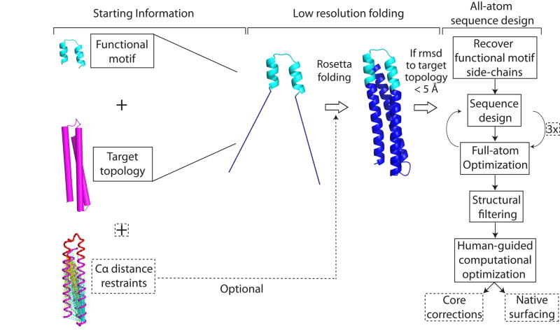
Vaccines prevent infectious disease largely by inducing protective neutralizing antibodies against vulnerable epitopes. Several major pathogens have resisted traditional vaccine development, although vulnerable epitopes targeted by neutralizing antibodies have been identified for several such cases. Hence, new vaccine design methods to induce epitope-specific neutralizing antibodies are needed. Here we show, with a neutralization epitope from respiratory syncytial virus, that computational protein design can generate small, thermally and conformationally stable protein scaffolds that accurately mimic the viral epitope structure and induce potent neutralizing antibodies. These scaffolds represent promising leads for the research and development of a human respiratory syncytial virus vaccine needed to protect infants, young children and the elderly. More generally, the results provide proof of principle for epitope-focused and scaffold-based vaccine design, and encourage the evaluation and further development of these strategies for a variety of other vaccine targets, including antigenically highly variable pathogens such as human immunodeficiency virus and influenza.


Figures

Extended Data Figure 1
Overview of the Fold from Loops (FFL) computational procedure. Initially large conformational spaces are sampled by low resolution folding, and subsequently iterative sequence design and small structural optimizations are performed to accommodate the target functional motif.
Extended Data Figure 2
Properties of designed proteins in this study. a) Sequence alignment of the FFL designs. 3LHP_S is the protein used as the template topology. b) Sequence alignment for the FFL_surf series designed based on the FFL_001 design model. c) Parameters and filtering criteria and results in the design process for FFL designs. a SD - standard deviation allowed on the constraints derived from the target topology; b Design epitope segment- design of residues within the epitope segment that were not part of the epitope-antibody interface; c Filtering criteria based on the helix bend angle; d Rosetta energy after human guided optimization. d) Structural diversity in the FFL design models. Values give the backbone RMSD in Å between two designs or between the template (3LHP_S) and the designs. e) Mutational diversity in the FFL designs. Values give the number of mutations between two designs or between the template (3LHP_S) and the designs.
Extended Data Figure 3
Structural properties of FFL designs in solution. a) Characterization of the oligomeric state by SECMALS. All molecules that showed a single monodisperse species by SEC had molecular weights computed from MALS that were consistent with expectation for a monomer (approximately 15 kDa). b) Secondary structure and thermal stability of FFL designs assessed by circular dichroism. Wavelength scans at T = 25 ° (left column) show the double minima typical for helical proteins. Thermal denaturation curves (right row) reveal high thermostability. d) HSQC spectra of several 15N labeled FFL designs. The spectra exhibit features typical for properly folded proteins with high alpha-helical content, particularly FFL_006. FFL_005 and FFL_007 exhibited reduced dispersion possibly due to self-association at higher concentrations.
Extended Data Figure 4
SPR data for FFL designs binding to Mota or Pali. a) Binding of FFL_005 and FFL_007 to Mota, in which the epitope scaffolds were amine-coupled to the sensor chip and Mota Fab was used as analyte. The concentrations of Mota Fab ranged from 950 nM to 436.5 pM and were used in serial dilutions with a dilution factor of three. b) Binding of FFL_001 and FFL_005 to Mota. Mota IgG was captured on the sensor chip by anti-human IgG and epitope scaffolds were used as analytes. The concentrations of scaffold ranged from 6.9 nM to 255.6 pM and were used in serial dilutions with a dilution factor of three. Kinetic fits are shown in red for both panels. c) Binding of FFL_001 and FFL_007 to Pali assessed by SPR. Pali IgG was captured by anti-human IgG on the sensor ship, and scaffolds were analytes. d) Mota binding specificity of FFL_001 assessed by SPR. Mota IgG was the ligand, captured by antihuman IgG on the sensor chip, and FFL_001 (blue) and an epitope point mutant of FFL_001 (FFL_001_K82E, black) were analytes at a concentration of 22 nM. The interaction between FFL_001 and Mota was eliminated by the point mutation.
Extended Data Figure 5
Crystallographic statistics for crystal structures determined. Values in parentheses refer to the highest resolution shell.
Extended Data Figure 6
Immunological evaluation of FFL scaffolds by different means. a) Evaluation of scaffolds as probes to detect the presence of epitope-specific antibodies in human sera. Sera from six healthy seropositive individuals were tested by ELISA for reactivity to FFL_001, FFL_001 with two different epitope point mutants (FFL_001_K82E and FFL_001_N72Y), and to recombinant RSV F glycoprotein. b) ELISA endpoint titers from mice immunized with immunogens shown on the x-axis. Autologous titers were measured against 001, 005, 007, or HBcAg particles without conjugated scaffold (triangles), and titers were also measured against RSV F protein (red). Titers after 2 immunizations are on the left, titers after 4 immunizations are on the right. c) ELISA endpoint titers for binding to recombinant RSV F protein, from NHPs immunized with 001, 005, 007, and HBcAg-FFL_001. d) RSV microneutralization assay results for NHPs immunized with 001, 005, 007, and HBcAg-FFL_001. In c) and d), values at each timepoint are mean ± standard deviation computed for the four animals per group at that timepoint.
Extended Data Figure 7
Neutralization of RSV by week 20, post-5 immunization NHP sera assessed by a flow cytometry based assay. a) The neutralization curves for several vaccinated animals are shown. 07C0012 was immunized with FFL_001; 07C004 and 07D039 were immunized with HBcAg-FFL_001; 07C0010 and 07D087 were immunized with FFL_007. b) Table showing 50% neutralization titers measured in two independent assays. c) Flow cytometry assay results for RSV subtypes A and B.
Extended Data Figure 8
Properties of NHP mAbs isolated by B cell sorting from an animal immunized with HBcAg-FFL_001. a) ELISA binding of recombinant NHP mAbs to FFL_001 (left) and recombinant RSV F glycoprotein (right). b) Sequence alignment of heavy (left) and light (right) chains of the Fv domains of NHP mAbs 17-HD9 and 31-HG7 along with Mota and Pali. c) SPR data for mAbs 17-HD9 and 31HG7 binding to FFL_001. mAb IgGs were captured by anti-human IgG on the sensor chip (mAbs were expressed with human Fc) and FFL_001 was flowed as analyte. d) Head-to-head comparison of the neutralization potency of NHP mAbs, Mota, and Pali in the plaque reduction assay. The data values are shown as mean ± standard deviation from two assays. The data were fit by the equation for one site specific binding with Hill slope, implemented in GraphpadPrism. According to the fits, the IC50s were 0.21 ug/mL (Pali), 0.046 ug/mL (Mota), 0.031 ug/mL (17-HD9), and 0.049 ug/mL (31-HG7). e) EC50 values for neutralization of RSV subtypes A and B by 17-HD9 and 31-HG7 as reported by the flow cytometry assay.
Extended Data Figure 9
SPR data for the binding of NHP mAbs to FFL_001 variants. a) FFL_001_surf1; b) FFL_001_K82E; c) FFL_001_R33C_N72Y_K82E. mAbs were captured by anti-human IgG on the sensor chip (mAbs were expressed with human Fc) and FFL_001 variants were flowed as analytes.
Extended Data Figure 10
Four complex structures of 17-HD9+peptide in the asymmetric unit, from PDB: 4N9G. The four complexes in the asymmetric unit consisted of two pairs of nearly identical structures (RMSD within each pair was 0.3 Å), with the pairs differing from each other primarily in the Fv angle of approach to the epitope (angle difference ∼9°) and in the Fab elbow angle (angle difference ∼10°); differences within the peptide between pairs were small (RMSD over peptide between pairs was 0.7 Å). a) Chains A+B+C; b) chains E+F+D; c) chains H+L+Y; d) chains M+N+Z. e) View of crystal packing interaction, in which the “backside” of one peptide interacts with the “backside” of another. Partial scaffolds (peptides) are packed against each other at crystal contacts between complexes through an interface outside of the epitope, with perfect dyad symmetry broken by a translation along the NCS dyad axis to accommodate complementary packing of apolar side-chains. The crystal packing is incompatible with the scaffold being present as a three helix bundle as in the Mota or 31-HG7 complex structures. Clear density was lacking for the scaffold outside the helix-turn-helix peptide. Scaffold missing density is possibly due to partial proteolysis or unfolding of the scaffold that may have occurred while purified Fab+scaffold complexes incubated at high concentration (∼10 mg/mL) in crystallization liquor for three months prior to crystal formation (see Supplementary Methods). The location and size of solvent channels in the crystal could accommodate the disordered region of the scaffold as an extended, flexible peptide unfolded under the conditions of crystallization, but it is also plausible that limited proteolysis has reduced the scaffold to a minimal structure protected by contacts with the antibody.
Figure 1
Figure 1. A novel computational method to design epitope-focused vaccines, illustrated with a neutralization epitope from RSV
Stages of computational design and immunological evaluation are shown; biophysical and structural evaluation are also important (see text).
Figure 2
Figure 2. Biophysical and structural characterization of scaffold FFL_001
a, Size exclusion chromatography coupled in-line with multi-angle light scattering measured a molecular weight in solution of ∼15 kDa, corresponding to a monomer. b, Circular dichroism data fit with a two state model showed that the protein had a melting temperature of 74 °C. Inset: The wavelength scan at 25 °C exhibited two minima characteristic of an all-helical protein,. c, Two dimensional 1H-15N HSQC spectrum at 25 °C and 600 MHz showed good peak dispersion typical of well-folded, alpha-helical proteins. ppm, parts per million. d, Surface plasmon resonance data and model fits (red lines) of the interaction with Mota Fab analyte, from which the dissociation constant (KD) was measured to be 29.9 pM. e, Crystal structure of unliganded FFL_005 (blue, green, and salmon helices, with yellow epitope), superimposed with the design model (gray with yellow epitope). f, Crystal structure of FFL_001 bound to Mota Fab, superimposed with the design model. Coloring as in e, but with the Fab light and heavy chains in gray or purple, respectively. g, Superposition of the epitope structure from unliganded FFL_005 (yellow) and the complex of peptide (green) bound to Mota from PDBid:3ixt. The positions of escape mutations for Pali (262 and 272) or Mota (272) are noted. h, Superposition of the Mota-liganded structures of FFL_001 and peptide (PDBid:3ixt). The antibody chains of 3ixt are colored in wheat, and the interfacial side-chains of both epitope and antibody are shown in stick representation.
Fig 3
Fig 3. Serological analysis of immunized macaques
a, ELISA endpoint titers measured against the autologous immunogen (left) or against RSV whole viral lysate (middle), and 50% neutralization titers as determined by the plaque reduction assay (right). The immunization groups are shown at far left, and the schedule is indicated at the bottom. Small symbols connected with dashed lines indicate individual animals. Large symbols connected with solid lines report group averages, with error bars showing standard deviations, measured over the four animals in each group at each time point. b, Comparison of 50% neutralization titers for sera from six RSV-seropositive humans and sera from eight macaques from weeks 12 and 20, measured side-by-side in the plaque reduction assay. Mean ± standard deviation for the human data are 218 ± 145. Two macaque data points at both week 12 and 20 are not visible in the graph because no neutralizing activity was detected. c, Comparison of 50% neutralization titers for sera from twenty RSV-seropositive humans and sera from five macaques from week 20, measured side-by-side in the flow cytometry assay. Mean ± standard deviation for the human data are 462 ± 792.
Fig 4
Fig 4. Analysis of monoclonal antibodies (mAbs) isolated from an immunized macaque
a, ELISA binding of the macaque mAbs (17-HD9 and 31-HG7) and Pali to RSV F. b, Neutralization of RSV by the macaque mAbs and Pali, measured by a microneuralization assay. The IC50s for Pali, 17-HD9 and 31-HG7 were 0.08, 0.005 and 0.007 μg/mL, respectively. c, MR model of 31-HG7 bound to FFL_001 (left), a crystal structure of 17-HD9 bound to a 35-residue helix-turn-helix peptide from FFL_001 (middle), and the crystal structure (PDB: 3ixt) of Mota bound to peptide. The three structures are aligned with respect to the helix-turn-helix epitope. d, Structural alignment of the helix-turn-helix epitopes bound to Mota (blue) and 17-HD9 (white), in which sidechains are colored orange if at least 15% of the total area (backbone+sidechain) of that residue is buried by the respective antibody. 9 positions are buried by both antibodies, two positions in the turn are buried only by 17-HD9 (P265 and T267, RSV residue numbering), and two positions near the peptide termini are buried only by Mota (S255 and N276). e, Close up view of the interface between 17-HD9 and helix-turn-helix epitope. Interaction residues are shown in stick, and the CDRH3 is colored red. K82/272 (scaffold numbering/RSV numbering), at the edge of the interface, is colored gray.

References

    1. Plotkin SA, Orenstein WA, Offit PA. Vaccines. 6. Elsevier; 2012.
    1. Rappuoli R, Mandl CW, Black S, De Gregorio E. Vaccines for the twentyfirst century society. Nat Rev Immunol. 2011;11:865–872. - PMC - PubMed
    1. Lozano R, et al. Global and regional mortality from 235 causes of death for 20 age groups in 1990 and 2010: a systematic analysis for the Global Burden of Disease Study 2010. Lancet. 2012;380:2095–2128. - PMC - PubMed
    1. Murray CJ, et al. Disability-adjusted life years (DALYs) for 291 diseases and injuries in 21 regions, 1990-2010: a systematic analysis for the Global Burden of Disease Study 2010. Lancet. 2012;380:2197–2223. - PubMed
    1. Rerks-Ngarm S, et al. Vaccination with ALVAC and AIDSVAX to prevent HIV-1 infection in Thailand. N Engl J Med. 2009;361:2209–2220. - PubMed

Publication types

MeSH terms