. 2014 Apr;24(2):108-19.
doi: 10.1097/CMR.0000000000000046.

Caveolin-1 is a risk factor for postsurgery metastasis in preclinical melanoma models

Affiliations

Caveolin-1 is a risk factor for postsurgery metastasis in preclinical melanoma models

Lorena Lobos-Gonzalez et al. Melanoma Res. 2014 Apr.

Abstract

Melanomas are highly lethal skin tumours that are frequently treated by surgical resection. However, the efficacy of such procedures is often limited by tumour recurrence and metastasis. Caveolin-1 (CAV1) has been attributed roles as a tumour suppressor, although in late-stage tumours, its presence is associated with enhanced metastasis. The expression of this protein in human melanoma development and particularly how the presence of CAV1 affects metastasis after surgery has not been defined. CAV1 expression in human melanocytes and melanomas increases with disease progression and is highest in metastatic melanomas. The effect of increased CAV1 expression can then be evaluated using B16F10 murine melanoma cells injected into syngenic immunocompetent C57BL/6 mice or human A375 melanoma cells injected into immunodeficient B6Rag1-/- mice. Augmented CAV1 expression suppresses tumour formation upon a subcutaneous injection, but enhances lung metastasis of cells injected into the tail vein in both models. A procedure was initially developed using B16F10 melanoma cells in C57BL/6 mice to mimic better the situation in patients undergoing surgery. Subcutaneous tumours of a defined size were removed surgically and local tumour recurrence and lung metastasis were evaluated after another 14 days. In this postsurgery setting, CAV1 presence in B16F10 melanomas favoured metastasis to the lung, although tumour suppression at the initial site was still evident. Similar results were obtained when evaluating A375 cells in B6Rag1-/- mice. These results implicate CAV1 expression in melanomas as a marker of poor prognosis for patients undergoing surgery as CAV1 expression promotes experimental lung metastasis in two different preclinical models.


Figures

Fig. 1
Fig. 1
CAV1 levels in human melanocytes and melanoma cell lines. Human melanocytes and melanoma cell lines were grown in 100 mm plates (see the Methods section). At 70% confluence, cells were harvested, extracts were prepared and proteins were separated by SDS-PAGE in 12% minigels (50 µg total protein per lane), transferred to nitrocellulose and analysed by western blotting with anti-CAV1 and antiactin antibodies. CAV1 protein levels were quantified by densitometric analysis. Numerical data were normalized to actin and averaged from three independent experiments (mean±SD, *P<0.05). (a, b) Representative blots are shown for the following: Hermes 3A and NOHM-4 (normal human melanocytes, M); MM200 (primary melanoma, PM), WM35 and WM1650 (radial growth phase, RGP); ME1402, Me10538, WM1341, WM98-1 and WM239A (vertical growth phase, VGP) and WM1158, COLO793 and DX3 cells (metastatic, Mts). Human fibroblasts were included as a positive control for CAV1 expression. (c) Relative CAV1 expression levels are graphed for the different human melanoma preparations. Statistically significant differences are indicated (*P<0.05). (d) Immunodetection of CAV1 in human samples using tissue arrays (normal tissue sample A04; MEL961 Pantomics). Individual melanocytes within the keratinocyte layer are highlighted (red arrows). Note that CAV1 was essentially undetectable in normal melanocytes when compared with keratinocytes (100×). Western blot insert: NhuM cells (melanocyte cell line) and HEKn (keratinocyte cell line) were compared. Note that CAV1 levels were at least 10-fold higher in HEKn cells. (e) CAV1 expression was determined as described for different stages of melanoma development. In the order of increasing invasiveness, representative samples of RGP, VGP and higher metastatic state are shown (E06, F11, H12; respectively; MEL961 Pantomics). CAV1, caveolin-1.
Fig. 2
Fig. 2
Tumour formation and lung metastasis of A375 (mock) and A375 (cav-1) cells. A375 (cav-1) and A375 (mock) cells were grown for 48 h in the presence of 1 mmol/l IPTG. Immunodeficient B6Rag1−/− mice were injected in the right flank with A375 (cav-1) or A375 (mock) cells (2.5×10⁶). (a) Schematic subcutaneous tumour formation. (b) Time course of tumour formation in B6Rag1−/− mice; black arrow corresponds to day 38. (c) Tumour volumes measured (day 38) for a total of five mice are shown. For CAV1-expressing cells and mock-transfected controls, tumour volumes were 268±232 and 1596±247 mm3, respectively (mean±SD). (d) Cell extracts from A375, A375 (mock) and A375 (cav-1) cells were analysed by western blotting with anti-CAV1 and antiactin antibodies. A representative western blot is shown. CAV1 protein levels observed in several experiments were quantified by scanning densitometry and normalized to actin (mean±SD, n=3; *P<0.05). A375 (mock) and A375 (cav-1) cells were injected intravenously (5×10⁶). Liver and lung tumour metastases were evaluated after 21 days. (e, f) Representative photographs of the liver (e) and lung (f) showing metastases by A375 (mock) and A375 (cav-1) cells (circles highlight tumours). (g, h) Quantification of total liver and lung mass evaluated 21 days after mice were injected with either A375 (mock) or A375 (cav-1) cells: liver 1.44±0.1 g compared with 2.73±0.4 g and lung 0.22±0.02 g compared with 0.32±0.02 g, respectively (*P<0.05). CAV1, caveolin-1; IPTG, isopropyl β-d-1-thiogalactopyranoside.
Fig. 3
Fig. 3
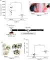
Tumour formation and lung metastasis of B16F10 (mock) and B16F10 (cav-1) cells. B16F10 (cav-1) and B16F10 (mock) cells were grown for 48 h in the presence of 1 mmol/l IPTG. C57BL/6 mice were injected simultaneously in the right flank with B16F10 (cav-1) and in the left flank with B16F10 (mock) cells (3×10⁵). (a) Tumour volumes measured (day 15) for a total of 14 mice. Means are shown. For CAV1-expressing cells and mock-transfected controls, tumour volumes were 460±81 and 1419±192 mm3, respectively (mean±SD). (b) Photograph of a necropsied mouse showing tumours formed in the left and right flank by B16F10 (mock) and B16F10 (cav-1) cells, respectively. (c) Schematic summarizing lung metastasis experiments. (d) Photographs showing lung metastasis by B16F10 (mock) and B16F10 (cav-1) cells. The black arrows indicate melanoma nodules. (e) C57BL/6 mice were injected with either 2×10⁵ B16F10 (cav-1) or B16F10 (mock) melanoma cells. Lung tumour mass was quantified after 21 days. Data from a total of 16 mice and the means are shown. Lung tumour mass for B16F10 (mock) and B16F10 (cav-1) cells was 9±2 and 28±2%, respectively (mean±SD). Statistically significant differences are indicated (***P<0.001). CAV1, caveolin-1; IPTG, isopropyl β-d-1-thiogalactopyranoside.
Fig. 4
Fig. 4
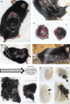
Melanoma surgery model. (a) C57BL6 mouse with a tumour of the bioethical permitted size (1500–1800 mm3). (b) Surgical removal of the tumour. (c) Surgically removed tumours of the same size derived from B16F10 (mock) (c1) and B16F10 (cav-1) cells (c2). (d) An animal immediately after surgery is shown with the open wound. (e) Operated animal, 2 days after surgery. (f) Following necropsy, 14 days after the initial surgery, recurrent tumours at the primary site were removed and volumes were measured. Tumours derived from B16F10 (mock) (f1) and B16F10 (cav-1) (f2) cells are shown. Note that tumours formed at the initial site after surgery by B16F10 (cav-1) cells were smaller than those obtained with B16F10 (mock) cells. (g) Lungs of mice killed 14 days after tumour surgery were analysed in animals initially injected subcutaneously with B16F10 (mock) (g1) or B16F10 (cav-1) (g2) cells. Note that lung metastasis was more prevalent for B16F10 (cav-1) cells.
Fig. 5
Fig. 5
CAV1 suppressed tumour recurrence and increased lung metastasis 14 days after surgery. (a) Schematic of events in surgery experiments; mice were injected on different days. Tumours between 1500 and 1800 mm3 in size were surgically removed. Animals were monitored after surgery and reincidence of tumour growth as well as lung metastasis were evaluated after sacrificing the animals 14 days later. (b) Tumours formed after surgery by B16F10 (cav-1) cells were smaller than those derived from B16F10 (mock) cells: 1280±80 and 2543±94 mm3, respectively. (c) Lung metastasis was significantly elevated for B16F10 (cav-1) compared with B16F10 (mock) cells: 20±5 and 7±2%, respectively. (d) Schematic of events in surgery experiments using mice injected the same day with B16F10 cells. Tumours between 1500 and 1800 mm3 in size were surgically removed. Reincidence of tumour growth as well as lung metastasis were evaluated after killing the animals 14 days later. (e) Tumours formed after surgery by B16F10 (cav-1) cells were smaller than those derived from B16F10 (mock) cells: 1200±98 and 2543±94 mm3, respectively. (f) Lung metastasis was significantly elevated for B16F10 (cav-1) compared with B16F10 (mock) cells: 19±4 and 6±2%, respectively. Statistically significant differences are indicated (***P<0.001, **P<0.01, *P<0.05). B16F10 (mock) and B16F10 (cav-1) cells were grown 48 h in the absence (−) or presence (+) of 1 mmol/l IPTG. Cell extracts were separated by SDS-PAGE and analysed by western blotting with anti-CAV1 and antiactin antibodies. Results from a representative western blot are shown. CAV1 protein levels quantified by scanning densitometry and normalized to actin (mean±SD, n=3; *P<0.05) are shown before injection of cells into animals (g). Tumours generated by B16F10 (mock) and B16F10 (cav-1) cells grown to the same final volume (1500–1800 mm3) (Fig. 4c1, c2) or grown to different volumes and excised the same day (Fig. 2b and c) were analysed for CAV1 protein levels. (h, i) Cells extracted from tumours in Fig. 2b and c were cultured for two passages (B16F10 extumour cells) and protein extracts were prepared. Statistically significant differences compared with B16F10 (mock) cells are indicated (*P<0.05). CAV1, caveolin-1; IPTG, isopropyl β-d-1-thiogalactopyranoside.
Fig. 6
Fig. 6
CAV1 suppressed tumour recurrence and increased lung metastasis of human melanomas after surgery in an mmunosuppressed mouse model. Immunodeficient B6Rag1−/− mice were injected into the right flank with either A375 (cav-1) or A375 (mock) cells (5×10⁶). (a) Schematic sequence of events in surgery experiments. Tumours between 800 and 1000 mm3 in size were surgically removed (white arrow). Animals were monitored after surgery and reincidence of tumour growth as well as lung metastasis were evaluated after killing the animals (black arrow in each case). (b, c) No tumours were detected after surgery for A375 (cav-1) cells. Alternatively, for A375 (mock) cells, mean tumour volumes of 697±310 mm3 were observed 50 days after surgery. Lung metastasis was significantly elevated after surgery for A375 (cav-1) compared with A375 (mock) cells. Quantification of total lung mass evaluated on the necropsy days (black arrow in b) (d) after mice were injected with either A375 (mock) or A375 (cav-1) cells: lung 0.275±0.05 g compared with 0.328±0.03 g, respectively (mean±SD), in the picture indicate the tumour mass in the case of A375 (cav-1) cells (*P<0.05). CAV1, caveolin-1.

Similar articles

Cited by

References

    1. Quest AF, Leyton L, Parraga M.Caveolins, caveolae, and lipid rafts in cellular transport, signaling, and disease.Biochem Cell Biol 2004;82:129–144 - PubMed
    1. Quest AF, Gutierrez-Pajares JL, Torres VA.Caveolin-1: an ambiguous partner in cell signalling and cancer.J Cell Mol Med 2008;12:1130–1150 - PMC - PubMed
    1. Quest AF, Lobos-Gonzalez L, Nunez S, Sanhueza C, Fernandez JG, Aguirre A, et al. The caveolin-1 connection to cell death and survival.Curr Mol Med 2013;13:266–281 - PubMed
    1. Lobos-Gonzalez L, Aguilar L, Diaz J, Diaz N, Urra H, Torres VA, et al. E-cadherin determines caveolin-1 tumor suppression or metastasis enhancing function in melanoma cells.Pigment Cell Melanoma Res 2013;26:555–570 - PMC - PubMed
    1. Nakashima H, Hamamura K, Houjou T, Taguchi R, Yamamoto N, Mitsudo K, et al. Overexpression of caveolin-1 in a human melanoma cell line results in dispersion of ganglioside GD3 from lipid rafts and alteration of leading edges, leading to attenuation of malignant properties.Cancer Sci 2007;98:512–520 - PMC - PubMed

Publication types