. 2014 Apr;5(2):217-30.
doi: 10.1007/s13244-014-0310-z. Epub 2014 Feb 7.

Non-contrast CT at comparable dose to an abdominal radiograph in patients with acute renal colic; impact of iterative reconstruction on image quality and diagnostic performance

Affiliations

Non-contrast CT at comparable dose to an abdominal radiograph in patients with acute renal colic; impact of iterative reconstruction on image quality and diagnostic performance

P D McLaughlin et al. Insights Imaging. 2014 Apr.

Abstract

Objectives: The aim was to assess the performance of low-dose non-contrast CT of the urinary tract (LD-CT) acquired at radiation exposures close to that of abdominal radiography using adaptive statistical iterative reconstruction (ASiR).

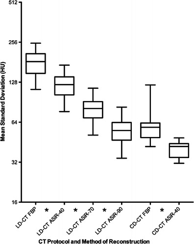
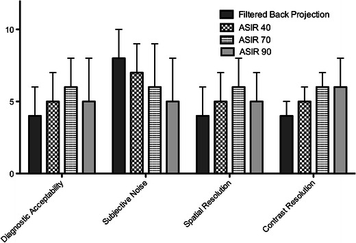
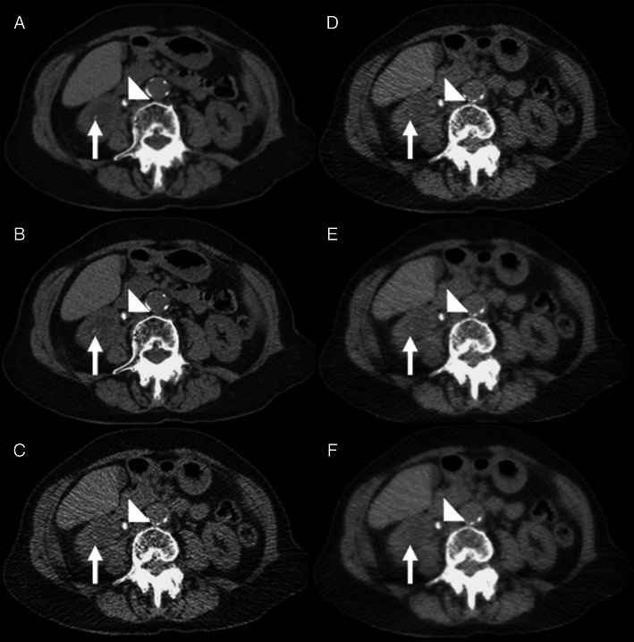
Methods: Thirty-three patients with clinically suspected renal colic were prospectively included. Conventional dose (CD-CT) and LD-CT data sets were contemporaneously acquired. LD-CT images were reconstructed with 40 %, 70 % and 90 % ASiR. Image quality was subjectively and objectively measured. Images were also clinically interpreted.

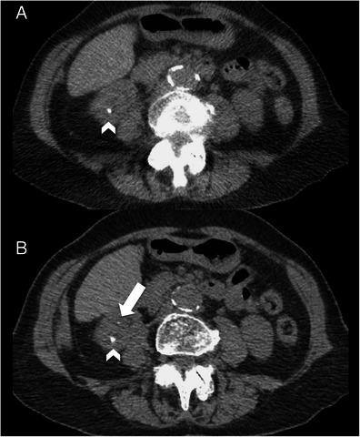
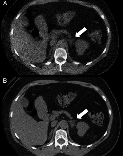
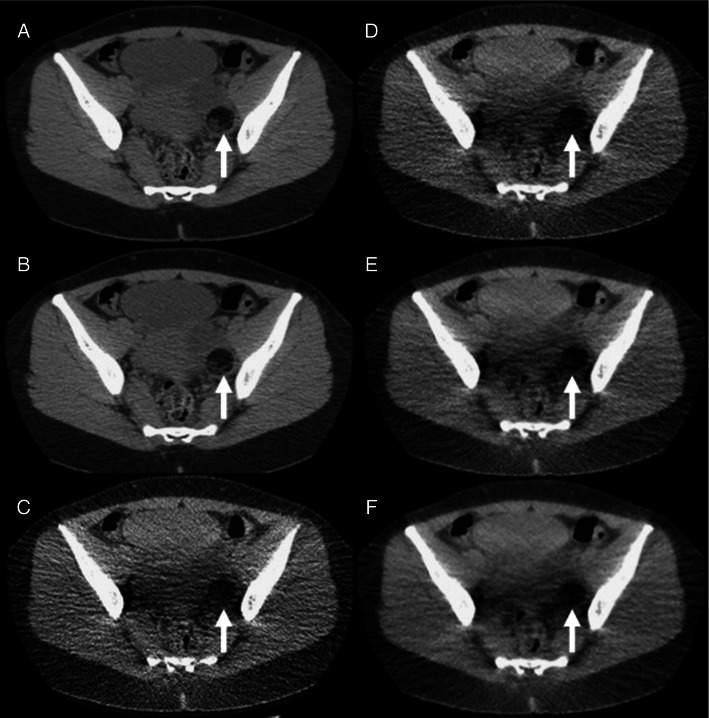
Results: Mean ED was 0.48 ± 0.07 mSv for LD-CT compared with 4.43 ± 3.14 mSv for CD-CT. Increasing the percentage ASiR resulted in a step-wise reduction in mean objective noise (p < 0.001 for all comparisons). Seventy % ASiR LD-CT images had higher diagnostic acceptability and spatial resolution than 90 % ASiR LD-CT images (p < 0.001). Twenty-seven calculi (diameter = 5.5 ± 1.7 mm), including all ureteric stones, were correctly identified using 70 % ASiR LD-CT with two false positives and 16 false negatives (diameter = 2.3 ± 0.7 mm) equating to a sensitivity and specificity of 72 % and 94 %. Seventy % ASiR LD-CT had a sensitivity and specificity of 87 % and 100 % for detection of calculi >3 mm.

Conclusion: Reconstruction of LD-CT images with 70 % ASiR resulted in superior image quality than FBP, 40 % ASIR and 90 % ASIR. LD-CT with ASIR demonstrates high sensitivity and specificity for detection of calculi >3 mm.

Teaching points: • Low-dose CT studies for urinary calculus detection were performed with a mean dose of 0.48 ± 0.07 mSv • Low-dose CT with 70 % ASiR detected calculi >3 mm with a sensitivity and specificity of 87 % and 100 % • Reconstruction with 70 % ASiR was superior to filtered back projection, 40 % ASiR and 90 % ASiR images.


Figures

Fig. 1
Fig. 1
Changes in mean objective noise (mean standard deviation of HU measured in 10 anatomical levels) encountered when 5-mm LD-CT images were reconstructed with FBP, and ASiR-40, ASiR-70, ASiR-90 and CD-CT images were reconstructed with FBP and ASiR-40 algorithms. (Star symbol denotes statistically significant difference, p < 0.05)
Fig. 2
Fig. 2
Changes in subjective image quality parameters (median score as measured at 5 anatomical levels) encountered when 5-mm LD-CT images were reconstructed with FBP, ASiR-40, ASiR-70 and ASiR-90. (All differences were statistically significant except comparison of contrast resolution scores between ASiR-70 and ASiR-90 images)
Fig. 3
Fig. 3
Changes in streak artefact score in the abdomen (light grey) and pelvic (dark grey) stations in patients with increasing BMI. 0, no streak artefact; 1, streak artefact present but not interfering with image interpretation; 2 streak artefact present and interfering with image interpretation
Fig. 4
Fig. 4
A 48-year-old female patient (BMI = 25.6) with acute right ureteric colic. a and b Axial conventional dose (5.1 mSv) non-contrast CT image reconstructed with 40 % ASiR (a) and 100 % FBP (b) showing an 8-mm right ureteric calculus (arrowhead) and an additional 2-mm renal calculus in the right lower pole (arrow) that was not prospectively detected on the low-dose images. c-f Axial low-dose (0.56 mSv) non-contrast CT image reconstructed with 100 % FBP (c), 40 % ASiR (d), 70 % ASiR (e) and 90 % ASiR (f) showing the obstructing 8-mm calculus in the proximal right ureter (arrowhead). The location of the smaller 2.1-mm calculus is also demonstrated (arrow). Note the ‘over smoothening’ of the soft tissues on the 90 % ASiR image compared to the other low-dose reconstructions
Fig. 5
Fig. 5
A 53-year-old female patient (BMI = 26.3) with acute left ureteric colic. a Axial low-dose (0.43 mSv) 2-mm non-contrast CT image reconstructed with 70 % ASiR showing a 5-mm calculus in the lower pole of the right kidney (arrowhead). b Axial conventional dose (5 mSv) 2-mm non-contrast CT image reconstructed with 40 % ASiR showing an additional 1.7-mm renal calculus in the right lower pole (arrow)
Fig. 6
Fig. 6
A 40-year-old female patient (BMI = 29.8) with suspected renal calculi. a Axial low-dose (0.45-mSv) 2-mm non-contrast CT image reconstructed with 70 % ASiR and b axial conventional dose (7 mSv) 2-mm non-contrast CT image reconstructed with 40 % ASiR showing a 3.2-cm low-density left adrenal mass consistent with a benign adenoma (arrow). This left adrenal mass was missed when viewed on the low-dose non-contrast CT images
Fig. 7
Fig. 7
A 29-year-old female patient (BMI = 20.8) with haematuria. a and b Axial conventional dose (3.6-mSv) 5-mm non-contrast CT images reconstructed with 40 % ASiR (a) and 100 % FBP (b) showing a 5.4-cm low-density left adnexal mass consistent with a dermoid cyst (arrow). This left adnexal mass was missed when viewed on the low-dose non-contrast CT images. c-f Axial low-dose (0.48-mSv) 5-mm non-contrast CT images reconstructed with 100 % FBP (c), 40 % ASiR (d), 70 % ASiR (e) and 90 % ASiR (f) showing the inferior detail of the left adnexal lesion relative to the conventional dose images (arrow). Again note the ‘over smoothening’ of the soft tissues on the 90 % ASiR image compared to the other low-dose reconstructions

References

    1. Smith RC, Rosenfield AT, Choe KA, Essenmacher KR, Verga M, Glickman MG, Lange RC. Acute flank pain: comparison of non-contrast-enhanced CT and intravenous urography. Radiology. 1995;194(3):789–794. doi: 10.1148/radiology.194.3.7862980. - DOI - PubMed
    1. Miller OF, Rineer SK, Reichard SR, Buckley RG, Donovan MS, Graham IR, Goff WB, Kane CJ. Prospective comparison of unenhanced spiral computed tomography and intravenous urogram in the evaluation of acute flank pain. Urology. 1998;52(6):982–987. doi: 10.1016/S0090-4295(98)00368-9. - DOI - PubMed
    1. Teichman JMH. Clinical practice. Acute renal colic from ureteral calculus. N Engl J Med. 2004;350(7):684–693. doi: 10.1056/NEJMcp030813. - DOI - PubMed
    1. Fielding JR, Steele G, Fox LA, Heller H, Loughlin KR. Spiral computerized tomography in the evaluation of acute flank pain: a replacement for excretory urography. J Urol. 1997;157(6):2071–2073. doi: 10.1016/S0022-5347(01)64676-7. - DOI - PubMed
    1. Smith RC, Verga M, McCarthy S, Rosenfield AT. Diagnosis of acute flank pain: value of unenhanced helical CT. AJR Am J Roentgenol. 1996;166(1):97–101. doi: 10.2214/ajr.166.1.8571915. - DOI - PubMed

LinkOut - more resources