. 2014 May;28(5):2047-61.
doi: 10.1096/fj.13-237388. Epub 2014 Feb 5.

Rapamycin nanoparticles target defective autophagy in muscular dystrophy to enhance both strength and cardiac function

Affiliations

Rapamycin nanoparticles target defective autophagy in muscular dystrophy to enhance both strength and cardiac function

Kristin P Bibee et al. FASEB J. 2014 May.

Abstract

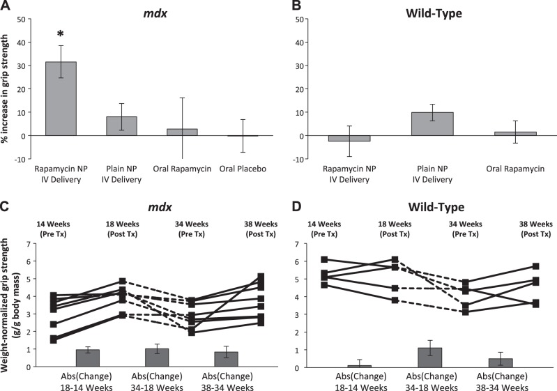
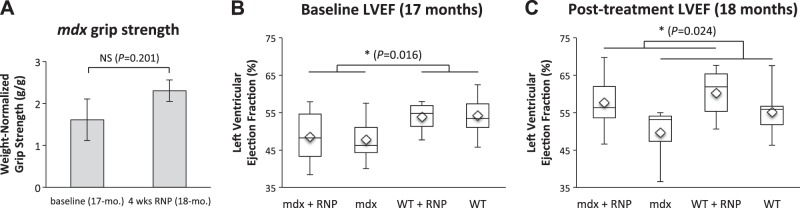
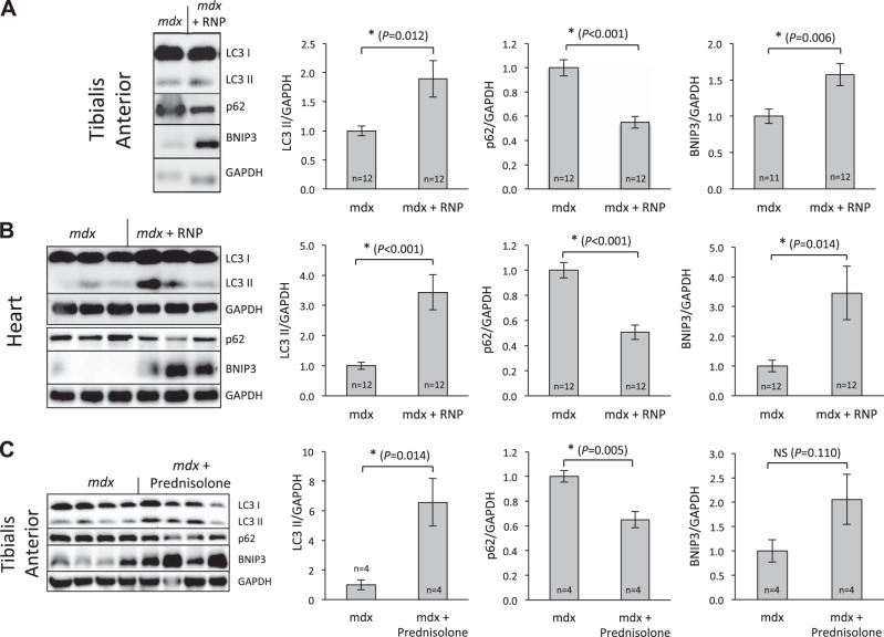
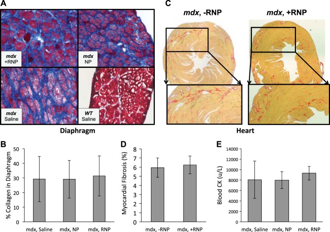
Duchenne muscular dystrophy in boys progresses rapidly to severe impairment of muscle function and death in the second or third decade of life. Current supportive therapy with corticosteroids results in a modest increase in strength as a consequence of a general reduction in inflammation, albeit with potential untoward long-term side effects and ultimate failure of the agent to maintain strength. Here, we demonstrate that alternative approaches that rescue defective autophagy in mdx mice, a model of Duchenne muscular dystrophy, with the use of rapamycin-loaded nanoparticles induce a reproducible increase in both skeletal muscle strength and cardiac contractile performance that is not achievable with conventional oral rapamycin, even in pharmacological doses. This increase in physical performance occurs in both young and adult mice, and, surprisingly, even in aged wild-type mice, which sets the stage for consideration of systemic therapies to facilitate improved cell function by autophagic disposal of toxic byproducts of cell death and regeneration.

Keywords: Duchenne; cardiomyopathy; drug delivery; nanomedicine; perfluorocarbon.


Figures

Figure 1.
Figure 1.
RNP treatment improves strength in mdx mice. A) Treatment with i.v. RNPs for 4 wk significantly increased mean weight-normalized grip strength in 14-wk-old mdx animals (n=16) vs. groups given plain NPs intravenously (n=18), equivalent oral doses of rapamycin (n=6), or oral placebo (n=6) (P=0.012, P=0.027, and P=0.016, respectively, using Fisher's protected least significant difference at 5% significance level). *P < 0.05. B) No significant difference in mean strength was observed for age-matched wild-type mice given either NPs (n=4) or RNPs intravenously (n=5) or oral rapamycin (n=6). C) Increase in weight-normalized strength in a subset of mdx mice (n=8) treated with i.v. RNPs occurred in both young (14–18 wk) and old (34–38 wk) animals (P=0.008 using a linear contrast model comparing pretreatment to posttreatment differences at a 5% significance level). Absolute values of the mean change in strength between time points are shown in columns below trend data. D) Wild-type mice (n=5) given similar treatment and drug holiday exhibit no significant difference between pretreatment and posttreatment strength (P=0.598). Bar graphs show mean ± sem values.
Figure 2.
Figure 2.
Aged mdx mice show muted responses in grip strength after treatment with RNP, but marked improvement in cardiac function commensurate with wild-type animals. A) Treatment of 17-mo-old mdx (n=8) for 4 wk yielded no significant increase in grip strength (P=0.201, 2-tailed paired t test). B) LVEF in wild-type mice is greater than mdx before treatment (P=0.016), as expected. C) Cardiac function significantly improved after RNP therapy, irrespective of animal type (16% difference for mdx and 9% difference for WT; P=0.024). NS, not significant. *P < 0.05.
Figure 3.
Figure 3.
NPs and rapamycin are effectively delivered to skeletal and cardiac muscle in mdx animals. A) 19F spectroscopy was performed on excised tissues to determine the levels of PFOB in mdx muscle. Each of the muscles surveyed showed NP (PFOB) uptake 24 h after last systemic injection. There was no significant difference in uptake between NPs and RNPs in any of the muscles tested (2-tailed t test, 5% significance level). B) mdx mice were given i.v. injections (1 ml/kg) of dual-labeled fluorescent NPs and euthanized 4 h postinjection following saline perfusion. Biceps (top panels), diaphragm, (middle panels), and gluteus (bottom panels) were excised, and adjacent sections were prepared for staining with H&E (left panels) and fluorescence microscopy (middle and right panels). NP lipid labeled with rhodamine is evident from detected fluorescence within skeletal muscle (middle panels), as is rapamycin within same tissue (right panels). C) NP lipid (rhodamine, displayed as blue) and rapamycin (Cy7.5, labeled as red) are colocalized in mdx cardiac tissue.
Figure 4.
Figure 4.
mdx animals exhibit a defect in autophagy that is restored by NP treatment. A) RNPs cause a decrease in S6 levels in mdx animals when compared to NP treatment. However, compared to saline treatment, the NPs increased S6 phosphorylation in all muscle groups tested, a trend not observed in the muscles from wild-type animals. B) Proteins were extracted from mdx and age-matched control animal muscle. Western blot analysis for p62 (n=5/group) and BNIP3 (n=3/group) reveal a defect in autophagy as mdx animals exhibited higher levels of p62 and lower BNIP3 when compared to wild-type (Student's t test at 5% significance level). Myosin was used as the loading control. C) RNA was isolated from the TA muscle of age matched mdx (n=5) and control mice (n=3). Quantitative PCR was performed and normalized to GAPDH. Graph demonstrates fold change in the following transcripts vs. control: there was no change in p62, BNIP3, or beclin. β2-Microglobin is increased as expected, because it is part of MHCI, which is known to be up-regulated in mdx muscle. Actin levels (normal) serve as an internal control. D) Representative Western blots demonstrate that both NPs and RNPs increase the levels of LC3B-II in mdx animals, whereas saline-treated animals exhibit a low level of LC3B-II expression, even when blocked with colchicine. Scatterplots show results from individual animals.
Figure 5.
Figure 5.
RNPs alter autophagic biomarkers in heart and skeletal muscle of mdx mice similar to that after steroid therapy. A) TA muscle in 17-mo-old mdx mice treated for 1 mo with RNPs exhibited significant effects of RNPs on p62 (decrease), BNIP3 (increase), and LC3II (increase). B) Cardiac data confirm that RNPs also enhance autophagy in the heart in the same manner. C) Young mdx mice given 42 wk of steroid therapy show similar autophagic responses in TA muscle. *P < 0.05.
Figure 6.
Figure 6.
Intravenously injected NPs do not affect short-term muscle cell destruction or fibrosis. A) Masson trichrome stains performed on diaphragm sections and visualized with light microscopy (×200) showed increased fibrosis (blue) for mdx animals relative to WT. B) No significant difference occurred over the short 4-wk test interval in mdx collagen levels after treatment with either RNPs, plain NPs, or saline (mixed model nested ANOVA, P=0.90). C, D) Picrosirius red stains of mdx mice (C) show diffuse heterogeneous fibrosis with no significant difference (P=0.664; D) when treated with RNPs. E) Serum CK levels for mdx mice exhibited no significant difference after treatment with either RNPs, NPs, or saline (ANOVA, P=0.71). Bar graphs show mean ± sd values.

References

    1. Hoffman E. P., Brown R. H., Jr., Kunkel L. M. (1987) Dystrophin: the protein product of the Duchenne muscular dystrophy locus. Cell 51, 919–928 - PubMed
    1. Moxley R. T., 3rd, Ashwal S., Pandya S., Connolly A., Florence J., Mathews K., Baumbach L., McDonald C., Sussman M., Wade C. (2005) Practice parameter: corticosteroid treatment of Duchenne dystrophy: report of the Quality Standards Subcommittee of the American Academy of Neurology and the Practice Committee of the Child Neurology Society. Neurology 64, 13–20 - PubMed
    1. Markham L. W., Spicer R. L., Khoury P. R., Wong B. L., Mathews K. D., Cripe L. H. (2005) Steroid therapy and cardiac function in Duchenne muscular dystrophy. Pediatr. Cardiol. 26, 768–771 - PubMed
    1. Biggar W. D., Harris V. A., Eliasoph L., Alman B. (2006) Long-term benefits of deflazacort treatment for boys with Duchenne muscular dystrophy in their second decade. Neuromuscul. Disord. 16, 249–255 - PubMed
    1. Balaban B., Matthews D. J., Clayton G. H., Carry T. (2005) Corticosteroid treatment and functional improvement in Duchenne muscular dystrophy: long-term effect. Am. J. Phys. Med. Rehab. 84, 843–850 - PubMed

Publication types

MeSH terms