. 2014 Apr;164(4):2167-83.
doi: 10.1104/pp.113.233585. Epub 2014 Feb 5.

Scavenging iron: a novel mechanism of plant immunity activation by microbial siderophores

Affiliations

Scavenging iron: a novel mechanism of plant immunity activation by microbial siderophores

Aude Aznar et al. Plant Physiol. 2014 Apr.

Abstract

Siderophores are specific ferric iron chelators synthesized by virtually all microorganisms in response to iron deficiency. We have previously shown that they promote infection by the phytopathogenic enterobacteria Dickeya dadantii and Erwinia amylovora. Siderophores also have the ability to activate plant immunity. We have used complete Arabidopsis transcriptome microarrays to investigate the global transcriptional modifications in roots and leaves of Arabidopsis (Arabidopsis thaliana) plants after leaf treatment with the siderophore deferrioxamine (DFO). Physiological relevance of these transcriptional modifications was validated experimentally. Immunity and heavy-metal homeostasis were the major processes affected by DFO. These two physiological responses could be activated by a synthetic iron chelator ethylenediamine-di(o-hydroxyphenylacetic) acid, indicating that siderophores eliciting activities rely on their strong iron-chelating capacity. DFO was able to protect Arabidopsis against the pathogenic bacterium Pseudomonas syringae pv tomato DC3000. Siderophore treatment caused local modifications of iron distribution in leaf cells visible by ferrocyanide and diaminobenzidine-H₂O₂ staining. Metal quantifications showed that DFO causes a transient iron and zinc uptake at the root level, which is presumably mediated by the metal transporter iron regulated transporter1 (IRT1). Defense gene expression and callose deposition in response to DFO were compromised in an irt1 mutant. Consistently, plant susceptibility to D. dadantii was increased in the irt1 mutant. Our work shows that iron scavenging is a unique mechanism of immunity activation in plants. It highlights the strong relationship between heavy-metal homeostasis and immunity.


Figures

Figure 1.
Figure 1.
Venn diagrams illustrating the transcriptome changes with DFO versus H2O. Comparison of DFO-up (A) and DFO-down (B) genes in leaves at indicated times after treatment.
Figure 2.
Figure 2.
Expression profiles of DFO-up genes in leaves and roots in response to DFO or CB siderophores. Leaves of Col-0 hydroponically grown plants were infiltrated with distilled water (control), 1 mm DFO, or 1 mm CB. Leaves and roots were harvested at indicated times after treatment. The expression of indicated genes was monitored by qRT-PCR using cDNA from leaves (A) or roots (B). Gene expression was normalized against synthetic constitutive gene transcript level (ACTIN [ACT] and ELONGATION FACTOR1α [EF1α]). Experiments were performed two times for DFO and three times for CB with similar results. Representative data are shown. Bars, sd of the normalized ratio. For each point, six plants were used, and three leaves per plant were infiltrated.
Figure 3.
Figure 3.
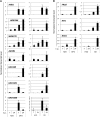
Overrepresented functional categories in up-regulated genes. A, Leaves at 7 and 24 hpi. B, Roots at 24 hpi. Data analysis was performed at Funcat (http://mips.helmholtz-muenchen.de/proj/funcatDB/). White bars indicate the percentage of category in the genome, and black bars indicate the percentage of the category in our dataset. P values are indicated for each category.
Figure 4.
Figure 4.
Expression profiles of DFO-up genes in leaves and roots in response to EDDHA treatment. Leaves of Col-0 hydroponically grown plants were infiltrated with 1 mm EDDHA or control solution (5 mm NaOH). Leaves and roots were harvested at indicated times after treatment. The expression of indicated genes was monitored by qRT-PCR from leaves (PR1, AtFER1, and bHLH100) or roots (IRT1 and NAS1). Gene expression was normalized against synthetic constitutive gene transcript level (ACT and EF1α). Experiments were performed three times with similar results. Representative data are shown. Bars, sd of the normalized ratio. For each point, six plants were used, and three leaves per plant were infiltrated.
Figure 5.
Figure 5.
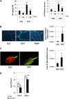
DFO triggers innate immune responses in Arabidopsis. Leaves of Col-0 hydroponically grown plants were infiltrated with indicated treatments. A, Leaves were harvested at 7 and 24 hpi, and total SA was quantified. JA amounts were expressed relative to the standard (“Materials and Methods”). DW, Dry weight. *P < 0.05 (Kruskal Wallis); bars, se. B, Picture and corresponding quantification of callose depositions detected with aniline blue staining in leaves 8 h after the indicated treatments. Bars = 200 µm. C, Picture and quantification of hydrogen peroxide staining in Arabidopsis leaves with the fluorescent dye 2',7'-dichlorfluorescein-diacetate 8 h after indicated treatments. *P < 0.05 (Mann and Whitney); bars, se. D, Virulence suppression activity of DFO in Arabidopsis. Col-0 leaves were infiltrated with DFO or water as control, which was followed by inoculation with the pathogen Pst DC3000 24 h later. Average bacterial counts per area (± se) are shown at indicated times after bacterial inoculation. **P < 0.01 (Mann and Whitney); bars, se. Experiments were performed three times at least with similar results. For each point, six plants were used, and three leaves per plant were infiltrated. [See online article for color version of this figure.]
Figure 6.
Figure 6.
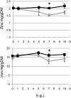
Time course of iron and zinc concentration in roots of DFO-treated plants. Plants were harvested at indicated times after 1 mm DFO (black) or water (gray) infiltration into leaves. Metal concentration was determined by inductively coupled plasma–atomic emission spectrometry on roots. DW, Dry weight. *P < 0.05 (Mann and Whitney); bars, se. Experiments were performed three times with similar results. For each point, six plants were used, and three leaves per plant were infiltrated.
Figure 7.
Figure 7.
Modification of iron distribution in leaf tissues in response to siderophores. Leaves of Col-0 hydroponically grown plants were infiltrated with 1 mm CB (B and E), 1 mm DFO (C and F), or distilled (A and D) water as control. Leaves were harvested at 5 hpi, and Perls’-DAB-H2O2 staining of thin sections was performed. D to F correspond to magnified zones of A to C, respectively. cw, Cell wall; n, nucleus; p, plastid. Bars in A to C = 20 µm. Bars in D to F = 5 µm.
Figure 8.
Figure 8.
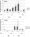
Role of protein dephosphorylation in iron homeostasis genes expression in response to DFO. Leaves of hydroponically grown plants were infiltrated with distilled water or 1 mm DFO. Leaves and roots were harvested at indicated times after treatment. Iron homeostasis gene expression was monitored by qRT-PCR using cDNA from leaves (bHLH100 and bHLH101) or roots (FRO2 and IRT1). Gene expression was normalized against synthetic constitutive gene transcript level (ACT and EF1α). OK, Okadaic acid. Experiments were performed three times with similar results. Representative data are shown. Bars, sd of the normalized ratio. For each point, six plants were used, and three leaves per plant were infiltrated.
Figure 9.
Figure 9.
The role of IRT1 in defense and iron homeostasis gene expression in response to DFO. A, Leaves of hydroponically grown plants were infiltrated with 1 mm DFO or water as control. Leaves and roots were harvested at indicated times after treatment. The expression of indicated genes was monitored by qRT-PCR from leaves (PR1, PDF1.2, AtFER1, and bHLH100) or roots (FRO2). Gene expression was normalized against synthetic constitutive gene transcript level (ACT and EF1α). Experiments were performed three times with similar results. Representative data are shown. Bars, sd of the normalized ratio; black bars, irt1-1 mutant; white bars, Ws. B, Pictures of callose depositions detected with aniline blue staining in leaves 8 h after the indicated treatments. Bars = 200 µm. C, Quantification of callose depositions in leaves 8 hpi **P < 0.01 (Mann and Whitney). Experiments were performed three times with similar results. [See online article for color version of this figure.]
Figure 10.
Figure 10.
Role of IRT1 in defense against D. dadantii. Leaves were inoculated with a D. dadantii bacterial suspension containing 107 cfu mL−1. The mean (± se; n = 30) of disease severity index scored at indicated times after inoculation is represented. Experiments were performed three times with similar results.
Figure 11.
Figure 11.
Model showing the responses of Arabidopsis to strong iron scavengers. A, Early events. Iron scavengers (EDDHA or siderophore) chelate iron in the surrounding tissue of the leaf and trigger an iron deficiency signal. This iron deficiency signal will be relayed by a signaling cascade, which potentially involves dephosphorylations and leads to the up-regulation of bHLH100 and bHLH101 in leaves and IRT1 and FRO2 in roots. B, Late events. Activation of IRT1 expression results in the uptake of iron and zinc at the root level, which is indicated by the thick orange arrow. This metal uptake is likely to cause a cellular stress in the treated leaves. The most important immunity and iron homeostasis markers are indicated. NON EXPRESSOR of PR1 is a protein involved in the SA signal transduction pathway shown to be required for PR1 activation by siderophores (Dellagi et al., 2009). Dotted arrows indicate that the activation is not direct. Genes up-regulated are surrounded by a colored box. Green and orange boxes refer to events occurring in leaves and roots, respectively. [See online article for color version of this figure.]

References

    1. Andrews SC, Robinson AK, Rodríguez-Quiñones F. (2003) Bacterial iron homeostasis. FEMS Microbiol Rev 27: 215–237 - PubMed
    1. Arnaud N, Murgia I, Boucherez J, Briat JF, Cellier F, Gaymard F. (2006) An iron-induced nitric oxide burst precedes ubiquitin-dependent protein degradation for Arabidopsis AtFer1 ferritin gene expression. J Biol Chem 281: 23579–23588 - PubMed
    1. Bakker PAHM, Pieterse CMJ, van Loon LC. (2007) Induced systemic resistance by fluorescent Pseudomonas spp. Phytopathology 97: 239–243 - PubMed
    1. Barberon M, Zelazny E, Robert S, Conejero G, Curie C, Friml J, Vert G. (2011) Monoubiquitin-dependent endocytosis of the iron-regulated transporter 1 (IRT1) transporter controls iron uptake in plants. Proc Natl Acad Sci USA 108: E450–E458 - PMC - PubMed
    1. Benning C. (2009) Mechanisms of lipid transport involved in organelle biogenesis in plant cells. In Annual Review of Cell and Developmental Biology, Vol 25 Annual Reviews, Palo Alto, CA, pp 71–91 - PubMed

Publication types

MeSH terms