. 2014 May;52(5):1330-7.
doi: 10.1128/JCM.03265-13. Epub 2014 Feb 5.

Universal influenza B virus genomic amplification facilitates sequencing, diagnostics, and reverse genetics

Affiliations

Universal influenza B virus genomic amplification facilitates sequencing, diagnostics, and reverse genetics

Bin Zhou et al. J Clin Microbiol. 2014 May.

Abstract

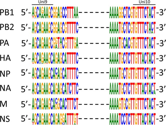
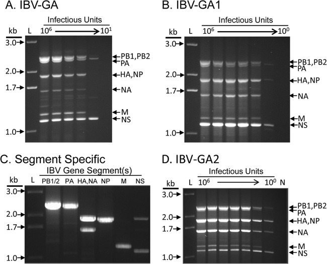
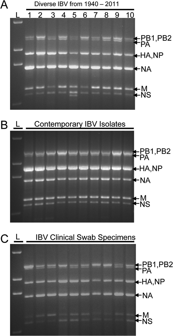
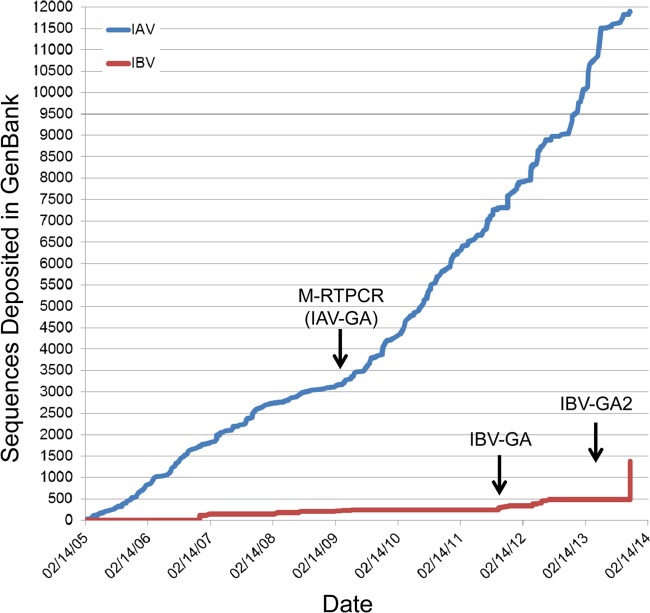
Although human influenza B virus (IBV) is a significant human pathogen, its great genetic diversity has limited our ability to universally amplify the entire genome for subsequent sequencing or vaccine production. The generation of sequence data via next-generation approaches and the rapid cloning of viral genes are critical for basic research, diagnostics, antiviral drugs, and vaccines to combat IBV. To overcome the difficulty of amplifying the diverse and ever-changing IBV genome, we developed and optimized techniques that amplify the complete segmented negative-sense RNA genome from any IBV strain in a single tube/well (IBV genomic amplification [IBV-GA]). Amplicons for >1,000 diverse IBV genomes from different sample types (e.g., clinical specimens) were generated and sequenced using this robust technology. These approaches are sensitive, robust, and sequence independent (i.e., universally amplify past, present, and future IBVs), which facilitates next-generation sequencing and advanced genomic diagnostics. Importantly, special terminal sequences engineered into the optimized IBV-GA2 products also enable ligation-free cloning to rapidly generate reverse-genetics plasmids, which can be used for the rescue of recombinant viruses and/or the creation of vaccine seed stock.


Figures

FIG 1
FIG 1
Conserved terminal sequences in the genome of IBVs. All the publicly available sequences (in cDNA sense) of IBVs were aligned for each gene segment. Nucleotides at the 20 terminal positions at each end of each segment were illustrated based on their relative frequencies at a specific position using the WebLogo application (16).
FIG 2
FIG 2
Efficiency of different primer sets in the amplification of IBV genome. The results of the three most efficient primer sets representing the three main stages of the development of the IBV genomic amplification technology are shown after agarose gel electrophoresis. RNA was extracted from 100 μl of 108 TCID50/ml of the B/Brisbane/60-10/2010 virus, eluted in 30 μl of nuclease-free water, and diluted in 1:10 series; 3 μl was used as a template for each RT-PCR. The equivalent amount of virus (TCID50) used in each RT-PCR is shown at the top of each lane. L, 1-kb Plus ladder (Life Technologies); N, negative control (no template). (A) IBV-GA. Three primers (Uni9/FluB1, Uni10/FluB1T, and Uni10/FluB1A) amplified the genome in the range of 106 to 101 TCID50/reaction. (B) IBV-GA2. Four primers (Uni9/FluB1G, Uni9/FluB1A, Uni10/FluB1T, and Uni10/FluB1A) amplified the genome in the range of 106 to 100 TCID50/reaction. (C) Segment-specific. Selected primer sets amplified different groups of genomic segments efficiently using 10 TCID50/reaction. Lanes, left to right: PB1/2-, PA-, HA/NA-, NP-, M-, and NS-specific primers (Table 1) were used. (D) IBV-GA2. Universal primer cocktail comprising the segment-specific primers used to amplify the genome in the range of 106 to 100 TCID50/reaction.
FIG 3
FIG 3
Universal IBV genomic amplification technology (IBV-GA2) amplifies genome of any IBV strain. L, 1-kb Plus ladder (Life Technologies). (A) Illustrates IBV genomes amplified from viruses isolated on different continents from 1940 to 2011. Lane 1, B/Lee/1940; lane 2, B/Russia/1969; lane 3, B/Hong Kong/5/1972; lane 4, B/Panama/45/1990; lane 5, B/Malaysia/11185/1996; lane 6, B/Victoria/143/2002; lane 7, B/England/145/2008; lane 8, B/Wisconsin/01/2010; lane 9, B/Hong Kong/259/2010; lane 10, B/Victoria/804/2011. IBVs in lanes 4 to 8 belong to the Yamagata lineage and viruses in lanes 9 and 10 belong to the Victoria lineage. (B) Representative example of the amplification of genomes from contemporary viruses isolated/cultured in Australia in 2011. (C) Representative example of the IBV-GA2 amplification of viral genomes directly from human swab specimens collected in the United States during the years of 2001 to 2006.
FIG 4
FIG 4
Recombination-based cloning of IBV genomic amplicons into reverse-genetics plasmids and the plaque phenotypes of rescued viruses. (A) Schematic diagram of the procedure used to clone genomic amplicons into the IBV recombination-based reverse-genetics plasmid pBZ66A12. To generate the reverse-genetics plasmid, a 22-bp fragment containing 9 bp that correspond to the 5′-termini of all vRNAs, followed by 4 bp (shown in italics) to create a PstI site, followed by 9 bp that correspond to the 3′-termini of all IBV vRNAs, was inserted between an RNA polymerase I promoter (pPolI) and terminator (Pol I-T). The genomic amplicons (1 segment shown) contain dsDNA copies of vRNA segments flanked by 10 (5′) or six (3′) bp that are derived from the 5′ tails incorporated into the primers (see examples in Table 1). The conserved Uni10 and Uni9 regions are shown in bold type. The genomic amplicons and PstI-linearized plasmid are mixed and treated with In-Fusion enzyme(s), and exonuclease activity exposes the complementary nucleotides at the termini leading to annealing and recombination (underlined nucleotides are part of the primer sequences used in the genomic amplification). (B) Plaque phenotypes of wtB/Russia/69 (left) and rB/Russia/69 (right). (C) Plaque phenotypes of wtB/Brisbane/60-10/2010 (left) and rB/Brisbane/60-10/2010 (right).
FIG 5
FIG 5
Complete influenza A and B virus genomes deposited in GenBank through the Influenza Genome Sequencing Project. Graph illustrates the number of full-length influenza A virus (IAV) (blue line) or influenza B virus (IBV) (red line) deposited in GenBank by the NIH/NIAID-sponsored Influenza Genome Sequencing Project since its initiation in 2005. Arrows indicate the initiation of universal genomic amplification procedures for influenza A virus (M-RTPCR) (19) or the influenza B virus (IBV-GA, IBV-GA2).

References

    1. WHO. 1980. A revision of the system of nomenclature for influenza viruses: a WHO memorandum. Bull. World Health Organ. 58:585–591 - PMC - PubMed
    1. Dowdle WR, Galphin JC, Coleman MT, Schild GC. 1974. A simple double immunodiffusion test for typing influenza viruses. Bull. World Health Organ. 51:213–215 - PMC - PubMed
    1. Feng L, Shay DK, Jiang Y, Zhou H, Chen X, Zheng Y, Jiang L, Zhang Q, Lin H, Wang S, Ying Y, Xu Y, Wang N, Feng Z, Viboud C, Yang W, Yu H. 2012. Influenza-associated mortality in temperate and subtropical Chinese cities, 2003–2008. Bull. World Health Organ. 90:279–288B. 10.2471/BLT.11.096958 - DOI - PMC - PubMed
    1. Jackson D, Elderfield RA, Barclay WS. 2011. Molecular studies of influenza B virus in the reverse genetics era. J. Gen. Virol. 92:1–17. 10.1099/vir.0.026187-0 - DOI - PubMed
    1. WHO. 2009. Influenza (seasonal) fact sheet no. 211. World Health Organization, Geneva, Switzerland: http://www.who.int/mediacentre/factsheets/fs211/en/

Publication types

LinkOut - more resources