. 2014 May;23(5):594-602.
doi: 10.1002/pro.2438. Epub 2014 Mar 11.

Structural characterization of MepB from Staphylococcus aureus reveals homology to endonucleases

Affiliations

Structural characterization of MepB from Staphylococcus aureus reveals homology to endonucleases

Sayeh Agah et al. Protein Sci. 2014 May.

Abstract

The MepRAB operon in Staphylococcus aureus has been identified to play a role in drug resistance. Although the functions of MepA and MepR are known, little information is available on the function of MepB. Here we report the X-ray structure of MepB to 2.1 Å revealing its structural similarity to the PD-(D/E)XK family of endonucleases. We further show that MepB binds DNA and RNA, with a higher affinity towards RNA and single stranded DNA than towards double stranded DNA. Notably, the PD-(D/E)XK catalytic active site residues are not conserved in MepB. MepB's association with a drug resistance operon suggests that it plays a role in responding to antimicrobials. This role is likely carried out through MepB's interactions with nucleic acids.

Keywords: DNA damage; PD-(D/E)XK endonucleases; antimicrobials; drug resistance.


Figures

Figure 1
Figure 1
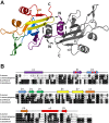
A: Structure of the MepB. The domain swapped N-terminal helices are shown encircled. One protomer is shown in gray, and the second is colored as a rainbow from the N terminus (purple) to the C terminus (red). B: Sequence alignment of MepB from S. aureus with homologs from Listeria monocytogenes, Bacillus Anthracis, and Clostridium botulinum. The secondary structure is shown over the alignment with the secondary structural elements in the same coloring scheme as Figure 1(A). Residues that align with the key catalytic residues for the PD-(D/E)XK superfamily are indicated with red circles.
Figure 2
Figure 2
Superposition of the MepB dimer (in orange) with two PvuII structures. A: Alignment of MepB a PvuII structure (in purple) that was determined in complex with DNA (PDB code 1PVI). B: Alignment of MepB a PvuII structure (in cyan) that was determined without DNA (PDB code 1PVU). The DNA is shown in green. All three structures were superimposed on the A molecule of the MepB dimer, which is shown encircled within the black oval. The DNA bound PvuII dimer is the most closed, and the MepB dimer is the most open.
Figure 3
Figure 3
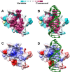
Surface representation of the MepB monomer structure. In (A) and (B) MepB is colored based on the conservation score obtained from the Consurf server (http://consurf.tau.ac.il/). In (C) and (D) MepB is colored based on its electrostatic potential as calculated by Chimera. In (B) and (D) the DNA from the I-Ssp6803I structure (PDB code 2OST) is overlaid on MepB, after the superposition of the two proteins.
Figure 4
Figure 4
Putative active site. Structure alignments of catalytic residues between MepB and I-Ssp6803I, and between MepB and PvuII. MepB, I-Ssp6803I, and PvuII are shown in orange, gray, and cyan, respectively. The three catalytic residues for I-Ssp6803I and PvuII are shown, along with the corresponding residues in MepB.
Figure 5
Figure 5
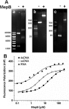
Binding assays. (A) Gel mobility shift assay. MepB binding to (a) 5 Kbp plasmid DNA on a 1% agarose gel, (b) ∼500 bp PCR product as seen on a 1% agarose gel, and (c) 26 bp oligonucleotide pair (ds26F) on a polyacrylamide gel. (B) Fluorescence polarization binding. Binding of MepB to (ds16F) is shown in black squares (▪), to (ss16F) is shown in open circles (o), and to (RNA16F) is shown in black triangles (▴). The MepB concentration is plotted on a logarithmic scale.

References

    1. Kaatz GW, Seo SM, O'Brien L, Wahiduzzaman M, Foster TJ. Evidence for the existence of a multidrug efflux transporter distinct from NorA in Staphylococcus aureus. Antimicrob Agents Chemother. 2000;44:1404–1406. - PMC - PubMed
    1. Kaatz GW, Moudgal VV, Seo SM. Identification and characterization of a novel efflux-related multidrug resistance phenotype in Staphylococcus aureus. J Antimicrob Chemother. 2002;50:833–838. - PubMed
    1. Kaatz GW, McAleese F, Seo SM. Multidrug resistance in Staphylococcus aureus due to overexpression of a novel multidrug and toxin extrusion (MATE) transport protein. Antimicrob Agents Chemother. 2005;49:1857–1864. - PMC - PubMed
    1. Huet AA, Raygada JL, Mendiratta K, Seo SM, Kaatz GW. Multidrug efflux pump overexpression in Staphylococcus aureus after single and multiple in vitro exposures to biocides and dyes. Microbiology. 2008;154:3144–3153. - PubMed
    1. Kaatz GW, DeMarco CE, Seo SM. MepR, a repressor of the Staphylococcus aureus MATE family multidrug efflux pump MepA, is a substrate-responsive regulatory protein. Antimicrob Agents Chemother. 2006;50:1276–1281. - PMC - PubMed

LinkOut - more resources