. 2014 Mar 4;111(9):3217-24.
doi: 10.1073/pnas.1400421111. Epub 2014 Feb 5.

Global analysis of cell cycle gene expression of the legume symbiont Sinorhizobium meliloti

Affiliations

Global analysis of cell cycle gene expression of the legume symbiont Sinorhizobium meliloti

Nicole J De Nisco et al. Proc Natl Acad Sci U S A. .

Abstract

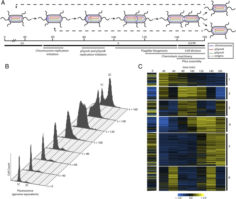
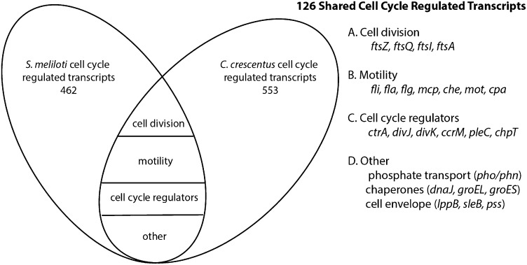
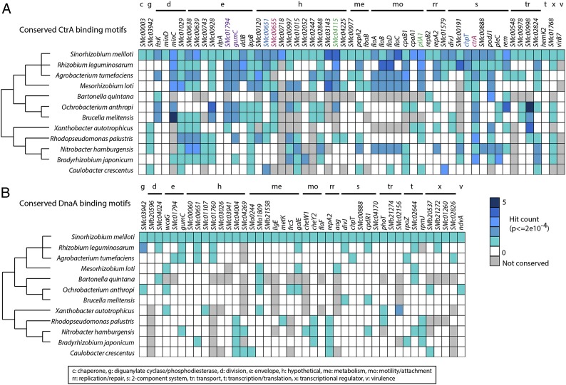
In α-proteobacteria, strict regulation of cell cycle progression is necessary for the specific cellular differentiation required for adaptation to diverse environmental niches. The symbiotic lifestyle of Sinorhizobium meliloti requires a drastic cellular differentiation that includes genome amplification. To achieve polyploidy, the S. meliloti cell cycle program must be altered to uncouple DNA replication from cell division. In the α-proteobacterium Caulobacter crescentus, cell cycle-regulated transcription plays an important role in the control of cell cycle progression but this has not been demonstrated in other α-proteobacteria. Here we describe a robust method for synchronizing cell growth that enabled global analysis of S. meliloti cell cycle-regulated gene expression. This analysis identified 462 genes with cell cycle-regulated transcripts, including several key cell cycle regulators, and genes involved in motility, attachment, and cell division. Only 28% of the 462 S. meliloti cell cycle-regulated genes were also transcriptionally cell cycle-regulated in C. crescentus. Furthermore, CtrA- and DnaA-binding motif analysis revealed little overlap between the cell cycle-dependent regulons of CtrA and DnaA in S. meliloti and C. crescentus. The predicted S. meliloti cell cycle regulon of CtrA, but not that of DnaA, was strongly conserved in more closely related α-proteobacteria with similar ecological niches as S. meliloti, suggesting that the CtrA cell cycle regulatory network may control functions of central importance to the specific lifestyles of α-proteobacteria.

Keywords: alpha-proteobacteria; cell cycle regulation; symbiosis.


Conflict of interest statement

The authors declare no conflict of interest.

Figures

Fig. 1.
Fig. 1.
Synchronization of S. meliloti and microarray analysis of cell cycle-regulated gene expression. (A) Illustration of S. meliloti cell cycle progression. The timing of cell cycle events is based on expression patterns of specific genes as described in results. S. meliloti is peritrichously flagellated and divides asymmetrically (23, 56). (B) FACS profiles from cell synchronization. At t = 0, 95% of cells are arrested in G1. At t = 60, replication has initiated and S phase starts. Cells have completed S phase at t = 140 and divide at t = 160. (C) Heatmap of the clustering of 462 cell cycle-regulated transcripts. Clusters are indicated on the right. Each row represents a single gene. The color scale bar corresponding to log-fold expression is included at the bottom of the heatmap.
Fig. 2.
Fig. 2.
Cell cycle gene expression of genes involved in various cellular processes. (A) Expression of genes involved in DNA processes (replication, repair, and segregation) in S. meliloti. Genes not included in our list of cell cycle-regulated transcripts are denoted by an asterisk (*). The color scale bar corresponding to log-fold expression is included at the bottom of the heatmaps. Values represent raw log fold change values. (B) Cell cycle expression patterns for genes involved in cell growth (ribosome and cell envelope) and division. (C) Cell cycle gene expression patterns of genes involved in motility and attachment including flagellar biosynthesis genes (and regulators), chemotaxis machinery, and genes required for pili biogenesis.
Fig. 3.
Fig. 3.
Expression profiles of conserved cell cycle regulators. Values depicted represent raw log fold change values. The color scale bar corresponds to log-fold expression change in comparison with unsynchronized culture. Genes not included in our list of cell cycle-regulated transcripts are denoted by an asterisk (*).
Fig. 4.
Fig. 4.
Comparison of genes demonstrating cell cycle-regulated transcription in S. meliloti and C. crescentus. Diagram of genes conserved between the S. meliloti and C. crescentus cell cycle-regulated datasets (18). One hundred twenty-six cell cycle-regulated genes were conserved between the two species and fell into the functional groups as illustrated in the Venn diagram.
Fig. 5.
Fig. 5.
Conservation of putative CtrA- and DnaA-binding sites in cell cycle-regulated genes. The upstream regulatory regions of genes with cell cycle-regulated transcripts were scanned for CtrA-and DnaA-binding motifs. “Hit count” signifies the number of significantly conserved full-length binding motifs that occur within the examined DNA segment. Homologs of genes containing hits from 11 related α-proteobacteria were also scanned for conserved CtrA and DnaA motifs. The tree to the left of the species’ names represents their evolutionary relationship as described in ref. . (A) S. meliloti cell cycle-regulated genes with the highest-scoring CtrA-binding motifs are displayed and the number of hits is denoted by different shades of blue as described in the legend. The presence of CtrA-binding sites in the homologous genes of related species is denoted in the same manner. Color-coded genes (i.e., pilA and SMc04115) share promoter regions. (B) S. meliloti cell cycle-regulated genes with the highest-scoring DnaA-binding motifs. The number of binding motifs and conservation of these motifs in other α-proteobacteria are denoted in the same manner as in A. A key explaining the putative function of the various genes displayed is provided at the bottom of the figure.

References

    1. Ettema TJ, Andersson SG. The alpha-proteobacteria: The Darwin finches of the bacterial world. Biol Lett. 2009;5(3):429–432. - PMC - PubMed
    1. Jones KM, Kobayashi H, Davies BW, Taga ME, Walker GC. How rhizobial symbionts invade plants: the Sinorhizobium-Medicago model. Nat Rev Microbiol. 2007;5(8):619–633. - PMC - PubMed
    1. Perret X, Staehelin C, Broughton WJ. Molecular basis of symbiotic promiscuity. Microbiol Mol Biol Rev. 2000;64(1):180–201. - PMC - PubMed
    1. Oldroyd GE. Speak, friend, and enter: Signalling systems that promote beneficial symbiotic associations in plants. Nat Rev Microbiol. 2013;11(4):252–263. - PubMed
    1. Mergaert P, et al. Eukaryotic control on bacterial cell cycle and differentiation in the Rhizobium-legume symbiosis. Proc Natl Acad Sci USA. 2006;103(13):5230–5235. - PMC - PubMed

Publication types

Associated data

LinkOut - more resources