. 2014 Feb 5;34(6):2025-36.
doi: 10.1523/JNEUROSCI.3736-13.2014.

Tuning to odor solubility and sorption pattern in olfactory epithelial responses

Affiliations

Tuning to odor solubility and sorption pattern in olfactory epithelial responses

John W Scott et al. J Neurosci. .

Abstract

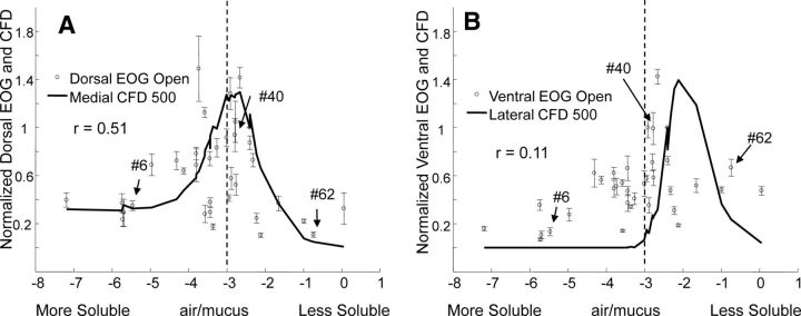
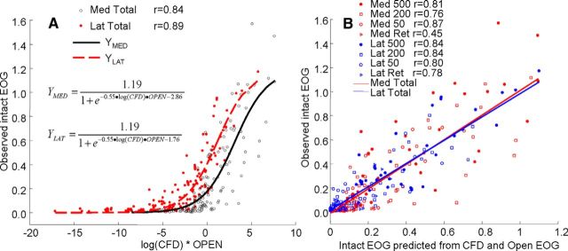
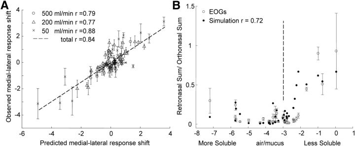
Odor information is first represented as a spatial activation pattern across the olfactory epithelium, when odor is drawn into the nose through breathing. This epithelial pattern likely results from both the intrinsic olfactory sensory neuron (OSN) sensitivity and the sorptive patterns imposed by the interaction of nasal aerodynamics with physiochemical properties of odorants, although the precise contributions of each are ill defined. Here, we used a computational fluid dynamics (CFD) model of rat nasal cavity to simulate the nasal aerodynamics and sorption patterns for a large number of odorants, and compared the results with the spatial neural activities measured by electro-olfactogram (EOG) under same flow conditions. The computational and experimental results both indicate greater sorption and response to a narrow range odorants as a function of their mucosal solubility, and this range can be further modulated by changes of intranasal flow rates and direction (orthonasal vs retronasal flow). A striking finding is that the profile of intrinsic EOG response measured in surgically opened nose without airflow constraints is similar to the shape of the sorption profile imposed by nasal airflow, strongly indicating a tuning process. As validation, combining the intrinsic response with the mucosal concentration estimated by CFD in nonlinear regression successfully accounts for the measured retronasal and orthonasal EOG response at all flow rates and positions. These observations redefine the role of sorption properties in olfaction and suggest that the peripheral olfactory system, especially the central zone, may be strategically arranged spatially to optimize its functionality, depending on the incoming stimuli.

Keywords: computational fluid dynamics; electro-olfactogram; olfactory sensory neuron; rat.


Figures

Figure 1.
Figure 1.
Computed tomography (CT) scans and the construction of the anatomical model. The full set of scans, consisting of 3300 images at an isotropic spatial resolution of 19 μm/pixel, were used to construct the anatomically accurate CFD model (see Materials and Methods). The sections on the right side are cross sections of the final CFD model at same location of example CT scans on the left. The lumen of the nasal cavity is shown in white. A, B, Arrows indicate the medial (A) and lateral (B) recording sites. The defects in the bone around those sites show how the bone was locally thinned to allow access to the epithelium.
Figure 2.
Figure 2.
Calculated odorant concentrations in olfactory mucosa under different flow conditions. A, The CFD calculations of odorant concentration for the three orthonasal flow rates and for the retronasal condition are shown in the plots on the left and right. The odorants are arrayed on the x-axis according to the log of air/mucus partition coefficients in Table 1. The values for heptanoic acid, 2-heptanone, and cyclohexane are indicated by arrows labeled with the numbers indicating their relative solubility in the mucosa (Table 1, Column 1). The vertical dotted lines corresponding to log(air/mucus) values of −3 help to indicate the shift in the distributions and the peaks between the two sites. The insets in the center of the panel illustrate the calculated intranasal concentrations in air for coronal sections corresponding to the positions of the medial electrodes at orthonasal 500 ml/min and incoming odorant concentration at the nostril of 100 ppm. The position of the medial electrodes is shown by the solid arrow. The position of the lateral electrodes was more caudal, but the position relative to the midline is shown by the dotted arrow. B, The upper row shows the absorbed concentrations in mucus (orthonasal 500 ml/min and nostril odorant concentration at 100 ppm) along the turbinate surface as viewed from the septum (top) and viewed from outside (bottom). The arrows at the top of each view in the upper row show the position of the medial electrode. The arrows at the top of each image in the lower row show the positions of the lateral electrodes for the intact preparation. The arrows at the right show the positions of four recording electrodes along the rostral surface of endoturbinate IV used in the open experiments. Rostral is to the left in both the medial and lateral views.
Figure 3.
Figure 3.
Responses and simulations in the intact preparation. A, B, EOGs for orthonasal flow at three flow rates (A) and for retronasal flow at the highest flow rate (B). The symbols indicate the response size relative to the orthonasal isoamyl acetate response at 500 ml/min ± SEM plotted against the log(air/mucus partition coefficient). The lines show the CFD simulation of odorant concentration in the epithelium at the recording sites under each flow condition. The CFD results are normalized to the CFD values for isoamyl acetate at 500 ml/min at the two sites. The vertical dotted lines, as in Figure 2A, help to emphasize the differences between the medial and lateral distributions. Note that the retronasal results are shown at an expanded vertical scale. The arrows in each plot point to the positions of the three odorants identified in Figure 2.
Figure 4.
Figure 4.
Comparison of smoothed EOG records and odorant concentrations. A, B, Intact medial and lateral EOG at orthonasal 500 ml/min compared with a moving average, where EOG values to odorants with similar mucosa solubility values (±0.5 log unit) are averaged. This average serves to remove the variabilities (neuronal, experimental, etc.) that are unrelated to sorption. C, D, The moving average response is compared with the CFD-calculated concentrations. In each case, the values are normalized to the value for isoamyl acetate. The vertical dotted lines corresponding to log(air/mucus) values of −3 help to compare the peaks with those in previous figures.
Figure 5.
Figure 5.
Responses in the open preparation and their comparison with intact responses. A, B, The mean responses ± SEM for the most dorsal and most ventral of the four electrodes in the open preparations for 35 of the odorants shown in Figure 1B. The solid line shows the CFD calculations of odorant concentration for the high flow rate scaled to isoamyl acetate response. The vertical dashed line at log(air/mucus) equals −3 helps indicate the shift in response distributions between the two plots. Odorants are identified as in Figure 1.
Figure 6.
Figure 6.
Prediction of intact EOG response by the regression on open response and calculated concentration. A, The open responses from Figure 4A and B were used as estimates of the inherent response of the olfactory epithelium to each odorant. These values were entered into a logistic regression equation with the logarithm of the calculated odorant concentration at each flow condition for the two intact sites to predict the intact response. The overall correlation including both sites and all four conditions is 0.86 (p < 0.001). B, The data of A are replotted to show that the relationship between predicted and observed values is generally linear. All the correlations in the figure are significant at p < 0.001 except that for the medial retronasal site for which p = 0.006.
Figure 7.
Figure 7.
Comparison of predicted response patterns and observed EOGs. A, Differences between the medial and lateral responses at the three orthonasal flow rates plotted against the prediction from the regression of Equation 1. B, Total (medial plus lateral) retronasal EOG response ± SEM normalized to total orthonasal response at the highest flow rate with the values predicted from total retronasal prediction from Equation 1 divided by the total orthonasal prediction. The vertical dotted line corresponding to log(air/mucus) values of −3 help to compare the retronasal sum with the values in previous figures.

Similar articles

Cited by

References

    1. Barlow R, Blake JF. Hill coefficients and the logistic equation. Trends Pharmacol Sci. 1989;10:440–441. doi: 10.1016/S0165-6147(89)80006-9. - DOI - PubMed
    1. Cenier T, McGann JP, Tsuno Y, Verhagen JV, Wachowiak M. Testing the sorption hypothesis in olfaction: a limited role for sniff strength in shaping primary odor representations during behavior. J Neurosci. 2013;33:79–92. doi: 10.1523/JNEUROSCI.4101-12.2013. - DOI - PMC - PubMed
    1. Cometto-Muñiz JE, Cain WS, Abraham MH. Quantification of chemical vapors in chemosensory research. Chem Senses. 2003;28:467–477. doi: 10.1093/chemse/28.6.467. - DOI - PubMed
    1. Coppola DM, Waggener CT, Radwani SM, Brooks DA. An electroolfactogram study of odor response patterns from the mouse olfactory epithelium with reference to receptor zones and odor sorptiveness. J Neurophysiol. 2013;109:2179–2191. doi: 10.1152/jn.00769.2012. - DOI - PubMed
    1. Curran-Everett D. Multiple comparisons: philosophies and illustrations. Am J Physiol Regul Integr Comp Physiol. 2000;279:R1–R8. - PubMed

Publication types

LinkOut - more resources