. 2014 Feb 5;34(6):2285-98.
doi: 10.1523/JNEUROSCI.4037-13.2014.

Phenotypic heterogeneity and plasticity of isocortical and hippocampal astrocytes in the human brain

Affiliations

Phenotypic heterogeneity and plasticity of isocortical and hippocampal astrocytes in the human brain

Alexander A Sosunov et al. J Neurosci. .

Abstract

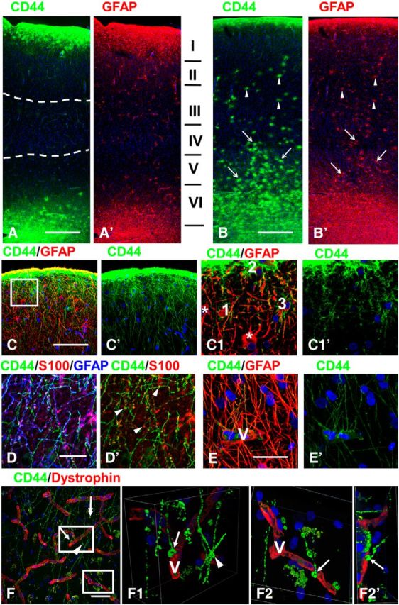
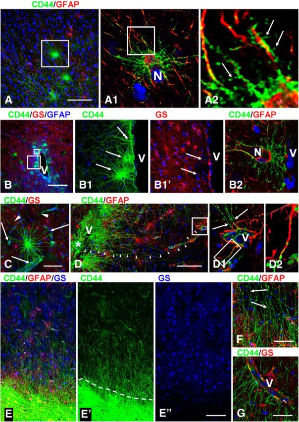
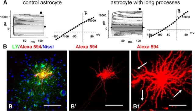
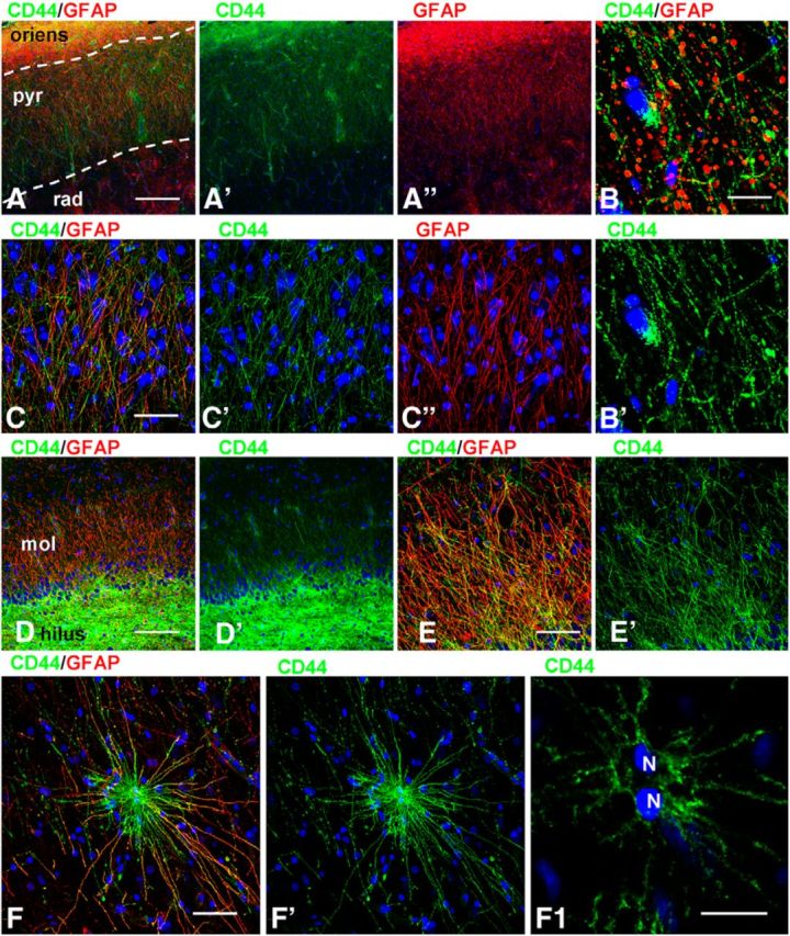
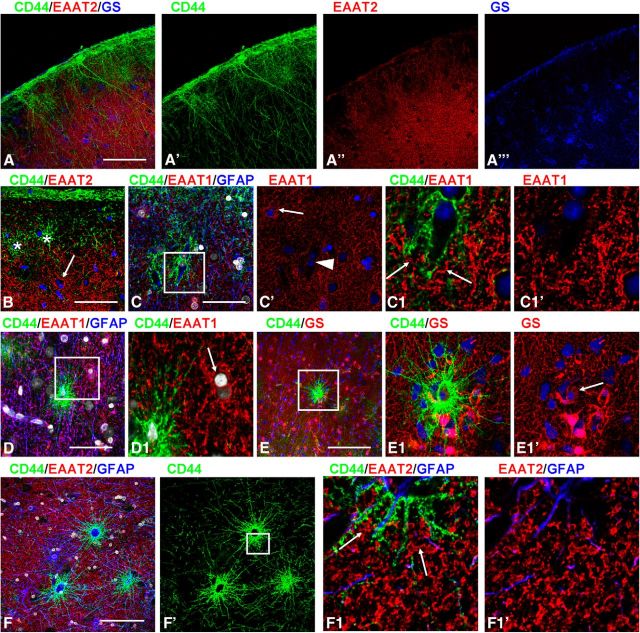
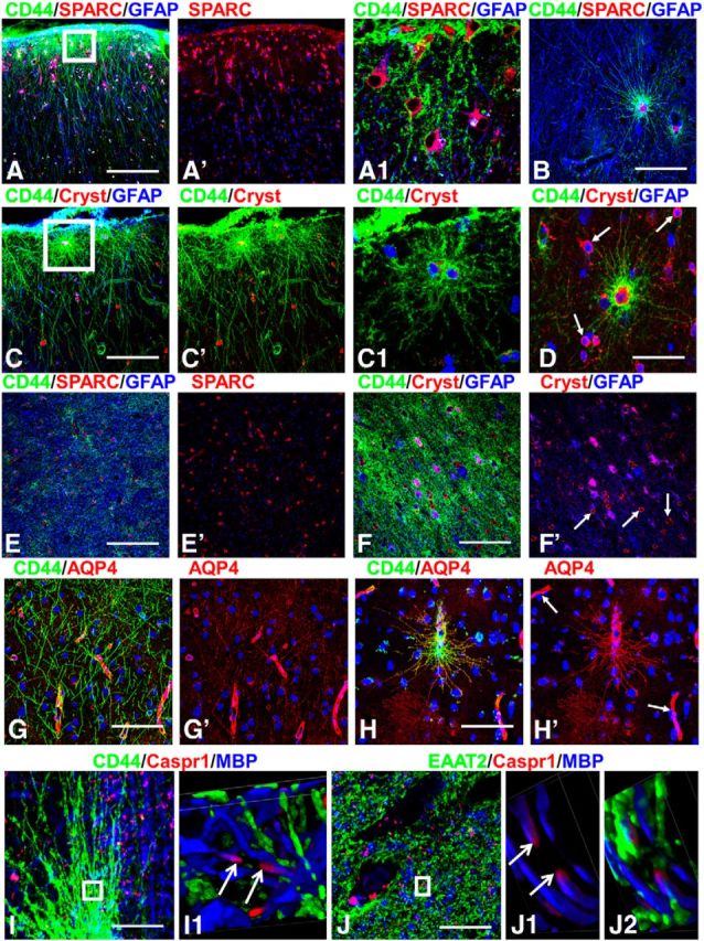
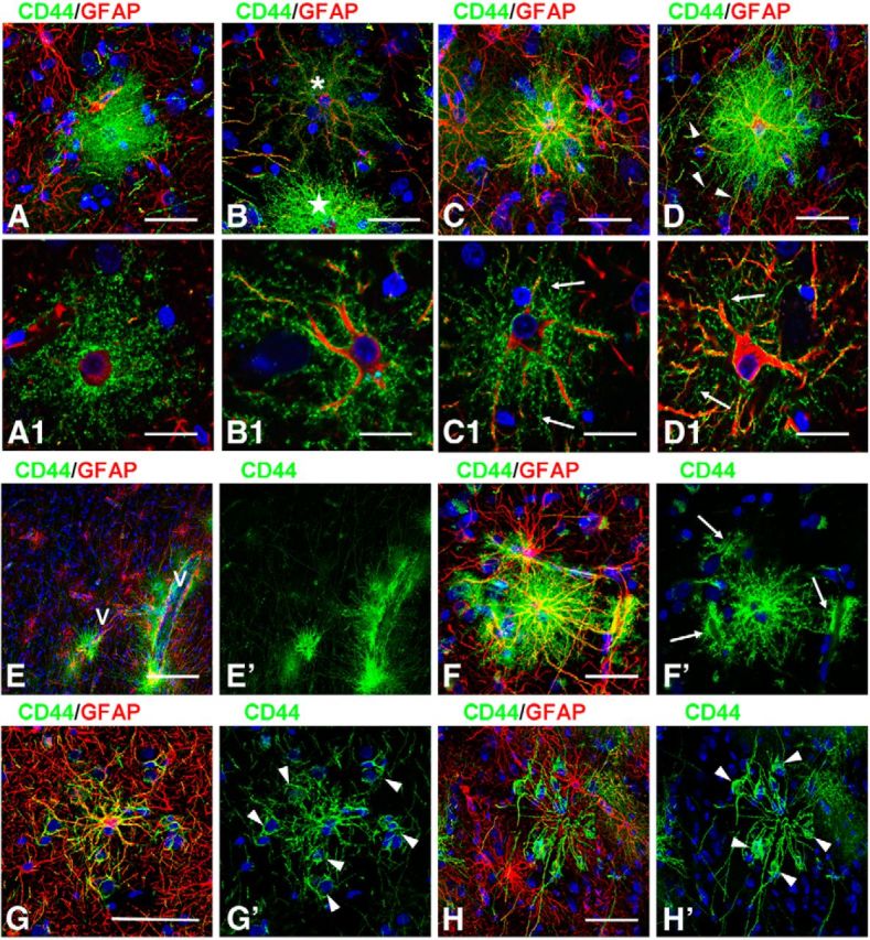
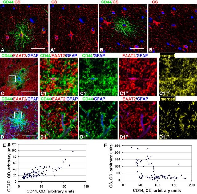
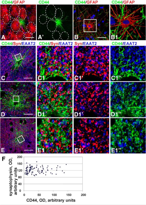
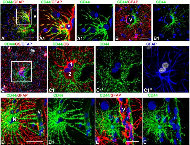
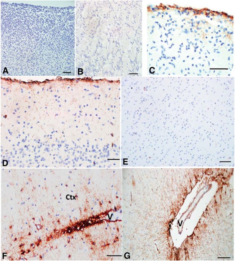
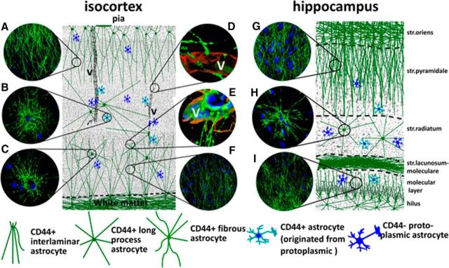
To examine the diversity of astrocytes in the human brain, we immunostained surgical specimens of temporal cortex and hippocampus and autopsy brains for CD44, a plasma membrane protein and extracellular matrix receptor. CD44 antibodies outline the details of astrocyte morphology to a degree not possible with glial fibrillary acidic protein (GFAP) antibodies. CD44+ astrocytes could be subdivided into two groups. First, CD44+ astrocytes with long processes were consistently found in the subpial area ("interlaminar" astrocytes), the deep isocortical layers, and the hippocampus. Many of these processes ended on blood vessels. Some were also found adjacent to large blood vessels, from which they extended long processes. We observed these CD44+, long-process astrocytes in every brain we examined, from fetal to adult. These astrocytes generally displayed high immunostaining for GFAP, S100β, and CD44, but low immunostaining for glutamine synthetase, excitatory amino-acid transporter 1 (EAAT1), and EAAT2. Aquaporin 4 (AQP4) appeared distributed all over the cell bodies and processes of the CD44+ astrocytes, while, in contrast, AQP4 localized to perivascular end feet in the CD44- protoplasmic astrocytes. Second, there were CD44+ astrocytes without long processes in the cortex. These were not present during gestation or at birth, and in adult brains varied substantially in number, shape, and immunohistochemical phenotype. Many of these displayed a "mixed" morphological and immunocytochemical phenotype between protoplasmic and fibrous astrocytes. We conclude that the diversity of astrocyte populations in the isocortex and archicortex in the human brain reflects both intrinsic and acquired phenotypes, the latter perhaps representing a shift from CD44- "protoplasmic" to CD44+ "fibrous"-like astrocytes.

Keywords: CD44; astrocyte; interlaminar astrocyte; protoplasmic astrocyte.


Figures

Figure 1.
Figure 1.
CD44+ astrocytes in human brain. A, B, Overview of two different specimens of temporal isocortex with few (A) or many (B) CD44+ astrocytes in the cortex. A, many long CD44+ astrocyte processes course through upper and lower cortical layers (dotted lines define their tips in mid-cortex). Note that their lengths in upper and lower cortex are similar. I–VI in A′ indicate cortical layers. B, Long-process (arrows) and short-process (arrowheads) CD44+ astrocytes occupy most cortical layers. Only some cells were indicated. C, Subpial interlaminar astrocytes. C1, Enlarged boxed area in C; single optical slices. Note (1) that CD44+ astrocytes (1–3) have only a few main branches with clearly outlined profiles, and (2) neighboring GFAP+ astrocytes (asterisks) are CD44 negative. D, Dense packing of long processes of subpial interlaminar astrocytes in layer II. Note that the majority of varicosities (arrowheads, marking only some varicosities) are immunopositive for GFAP and S100β. E, Many long processes of subpial CD44+ astrocytes end on a blood vessel (V) in cortical layer II. F, Endings of long-process CD44+ astrocytes in the lower part of layer III. One CD44+ process ends with an enlargement in the neuropil (two-headed arrow). F1, 2D views of a 3D reconstruction of enlarged upper boxed area in F showing end foot (arrow in F and F1) at a vessel wall (V). Note that the varicose-like enlargement that looks like a “free ending” (arrowhead) leaves the volume of the optical image z-stack. F2, F2, 2D views shown at different angles of a 3D reconstruction of enlarged lower boxed area in F. V, Blood vessel visualized with dystrophin. Confocal microscopy. Scale bars: A, B, 450 μm; C, 115 μm; D, E, 35 μm; F, 50 μm.
Figure 2.
Figure 2.
CD44+ astrocytes with long processes in temporal isocortex. A, Long-process CD44+ astrocytes in the deep cortical layers. A1, Enlarged boxed area in A; single optical slice. Note perineuronal (N) position of CD44+ astrocyte. A2, Enlarged boxed area in A1. The main branches have only small spine-like protrusions (arrows) delineated with CD44. B, CD44+ astrocytes with long processes near a large blood vessel (V) in cortical layer IV. B1, Enlarged lower boxed area in B. Pedivascular astrocytes (arrows) in B1 have low levels of GS; double-headed arrow indicates perivascular astrocyte with long processes. B2, Enlarged upper boxed area. The astrocyte that occupies a perineuronal (N) position sends several end feet to blood vessel (V). C, Astrocyte in layer IV sends end feet to several blood vessels (arrows), whereas other processes pass over these vessels (arrowheads) and end beyond the area of the image. D, A long process (marked with arrowheads) of a perivascular (V) astrocyte (asterisk) ends on small blood vessels (see in D1, enlarged boxed area in D), Note in D1 that several CD44+ processes (arrows) going from upper parts of cortex make end feet on the same vessel (V). D2, Enlarged boxed area in D1 shows that the long CD44+ process “crawls” along a CD44−/GFAP+ astrocyte process approaching the vessel. CD44+ and GFAP+ processes are marked with green and red arrowheads, respectively, before they touch each other. E, Long processes ascending from white matter astrocytes into lower cortical layers. The gray matter–white matter border is defined with a dotted line (E) and is clearly discriminated by CD44 and GS immunostaining. F, Many thin astrocyte processes in lower cortical layers do not show detectable levels of GFAP immunostaining (arrows, marking only some processes). G, Many long astrocyte processes in the lower cortical layers end on blood vessels (V). Confocal microscopy. Scale bars: A, 180 μm; B, 215 μm; C, D, 120 μm; E, 85 μm; F, 40 μm; G, 30 μm.
Figure 3.
Figure 3.
A, Representative current traces were evoked by a step voltage protocol (from −160 to +60 mV in 10 mV increments; 100 ms duration). The current–voltage (I–V) curves were obtained from the measurements near the end of each voltage step (small black squares above current traces). Note the absence of difference between control (short processed astrocyte, left) and astrocyte with long processes (right). B, A subpial interlaminar astrocyte was filled with Lucifer yellow (for intercellular coupling) and with Alexa Fluor 594 (which does not penetrate gap junctions). Many neighboring astrocytes were filled with Lucifer yellow (B). Cell processes of the injected astrocyte have short spine-like protrusions (arrows in B1, enlargement of B). Confocal microscopy. Scale bars: B, B, 125 μm; B1, 25 μm.
Figure 4.
Figure 4.
CD44+ astrocytes in hippocampus. A, C, A dense net of long astrocyte processes covers the pyramidal layer in CA1. A, Dotted lines outline stratum pyramidale (pyr): surgical tissue. B, Many large varicosities in long astrocyte processes in stratum pyramidale: autopsy tissue. D, E, CD44+ processes cover the molecular layer of the dentate gyrus (mol). F, A CD44+ astrocyte with long processes in the CA1 stratum radiatum. Note that this astrocyte has two nuclei (N) (F1, enlargement of F; single optical slice). oriens, Stratum oriens; rad, stratum radiale. Confocal microscopy. Scale bars: A, 210 μm; B, 25 μm; C, D, 120 μm; E, 35 μm; F, 30 μm; F1, 15 μm.
Figure 5.
Figure 5.
CD44+ astrocytes with long processes display a phenotype similar to that of fibrous astrocytes. A, EAAT2 (red) and GS (blue) are low in subpial CD44+ (green) astrocytes. B, Subpial CD44+ astrocytes (asterisks) display much less EAAT2 immunolabeling than neighboring CD44− astrocyte (arrow). Single optical slice. C, EAAT1 signal is low in subpial CD44+ astrocytes with long processes. Compare CD44+astrocyte (arrowhead) and CD44− astrocyte (arrow). C, Single optical slice. C1, Enlarged boxed area in C. Blue: C, GFAP; C, C1, C1, nuclei. Note that EAAT1+ profiles (arrows) are located in the immediate vicinity of the CD44+ astrocyte cell body. D, Low level of EAAT1 immunolabeling of a CD44+ astrocyte with long processes in cortical layer V. D1, Enlarged boxed area in D, single optical slice, protoplasmic EAAT1+/CD44− astrocyte marked with arrow. E, F, Levels of GS (E) and EAAT2 (F) are low in CD44+ astrocytes with long processes in deep cortical layers. E1, F1, Enlarged boxed areas in E and F, respectively. E1, CD44+ astrocyte is marked with arrow. F1, Note that EAAT2+ processes (arrows) of an adjacent CD44− astrocyte are located near the cell body of the CD44+ astrocyte and in between the CD44+ processes. Confocal microscopy. Scale bars: A, 110 μm; B, 55 μm; C, 35 μm; D, 110 μm; E, 175 μm; F, 130 μm.
Figure 6.
Figure 6.
A–D, SPARC (A, B) and αB-crystallin (Cryst; C, D) are present in long-process astrocytes, shown in subpial (A, C) and in deep cortical layers (B, D). Cryst+ oligodendrocytes in D are marked with arrows. A1, C1, Enlarged boxed areas from A and C, respectively. SPARC (E) and αB-crystallin (F) in subcortical white matter. Arrows in F mark oligodendrocytes. G, H, AQP4 is observed in all processes in CD44+ astrocytes. G, Long processes of interlaminar astrocytes are AQP4+. H, Long-process CD44+ astrocyte near blood vessel displays immunoreactivity for AQP4 in processes. Note high levels of AQP4 near blood vessels (arrows). I, CD44+ long process astrocytes do not contact nodes of Ranvier visualized as the unstained part of the axon (depicted with arrows in I1) between two Caspr1+ ends of neighboring myelin sheaths. I1, 2D views of 3D reconstruction of enlarged boxed area in I. J, Processes of protoplasmic astrocytes visualized with EAAT2 located close to a node. J1, J2, 2D views of 3D reconstruction of enlarged boxed area in J. Note that EAAT2+ (green)-immunopositive processes are near the node (defined as space between arrows). Confocal microscopy. Scale bars: A, B, 130 μm; C, 100 μm; D, 55 μm; E, 130 μm; F, 130 μm; G, 85 μm; H, 120 μm.
Figure 7.
Figure 7.
Variability in the morphologies of CD44+ cortical astrocytes without long processes. A, A bushy-like protoplasmic astrocyte. B, Astrocyte (asterisk) with spongioform-like main processes with only few secondary and tertiary branches. Note CD44+ astrocyte (star) with many processes nearby. C, D, CD44+ astrocytes with reduced numbers of miniature leaf-like processes. Note the abundance of thin processes originated from perikarya in C1 and D1. Several thin, long processes (D, arrowheads) extend beyond the dense proximal processes. A1, B1, C1, and D1 show enlargements of A and B (top astrocyte), C, and D, respectively; single optical slices. E, CD44+ astrocytes with long and short processes residing near large blood vessels (V) in cortical layer IV. Note the many long CD44+ astrocyte processes passing through the neuropil. F, CD44+ protoplasmic-like astrocyte with end feet on three separate blood vessels (arrows in F). Note that end feet occupy large areas of blood vessel walls; single optical slice in F. G, H, Processes of CD44+ astrocytes making contacts around neighboring neurons (arrowheads, neurons identified with Nissl staining used in the blue channel). Note that processes lack miniature processes. Confocal microscopy. Scale bars: A–C, D, 40 μm; A1, B1, C1, D1, 25 μm; E, 145 μm; F, 55 μm; G, H, 45 μm.
Figure 8.
Figure 8.
Variability in immunohistochemical properties of CD44+ cortical astrocytes without long processes. A, B, Different levels of GS in CD44+ astrocytes. C, D, Moderate (C) and severe (D) decrease in EAAT2 immunolabeling in CD44+ astrocytes. C1, D1, Enlarged boxed areas in C and D, respectively. C1‴ and D1‴, Areas of CD44 and EAAT2 colocalization (yellow): note the greater amount of overlap in C1‴ than in D1‴. Confocal microscopy. Scale bars: A, B, 60 μm; C, 100 μm; D, 95 μm. E, Optical density reading of GFAP and CD44 in CD44+ astrocytes in mid-cortex. Note the significant direct correlation: r = 0.619, p < 0.001. F, Optical density reading of GS and CD44 in CD44+ astrocytes in mid-cortex. Note the significant inverse correlation: r = 0.456, p < 0.001.
Figure 9.
Figure 9.
Protoplasmic astrocyte domain organization is violated by CD44+ astrocytes. A, Long CD44+ processes pass through the domains (dotted lines) of neighboring CD44− astrocytes. B, Many CD44+ long processes (green) pass through the processes of a CD44− astrocyte. B1, 2D image of 3D reconstruction of enlarged boxed area in B. Note that CD44+ processes pass in close vicinity to the cell body of a CD44−/GFAP+ astrocyte. C–E, Distribution of synaptophysin immunopositive puncta in the vicinity of CD44+ astrocytes with long (C) and short (D, E) processes. C1, D1, E1, Enlarged boxed areas in C–E, respectively; single optical slices. Note that there is less CD44 immunostaining in C than in D and E. Confocal microscopy. Scale bars: A, 60 μm; B, 45 μm; CE, 55 μm. F, Optical density reading of synaptophysin puncta and CD44 in CD44− and CD44+ astrocytes without long processes in mid-cortex. Note the absence of correlation, r = 0.0819, p < 0.001.
Figure 10.
Figure 10.
Microheterogeneity in astrocyte properties. A, Two neighboring perivascular astrocytes differ in CD44 levels. A1, Enlarged boxed area in A. Note that the astrocyte marked with an asterisk is CD44+, whereas the other astrocyte (#) is CD44−. B, CD44+astrocyte covers only half of the perimeter of a blood vessel (V). C, Two astrocytes (doublet) differ in CD44 levels and display different levels of GS and GFAP. C1, Enlarged boxed area in C. The numbers 1 and 2 indicate two astrocytes with different immunohistochemical phenotypes. D, E, CD44+ long-process astrocyte (D) and CD44+short-process astrocyte (E) have different end feet. Note that end feet of the protoplasmic (short-process) astrocyte occupy much more vascular surface area. Note also in these images the different CD44+ and GFAP+ processes. Confocal microscopy. Scale bars: A, 75 μm; B, 90 μm; C, 55 μm; D, E, 85 μm.
Figure 11.
Figure 11.
CD44+ astrocytes in fetal and young CNS. A, B, At 19 weeks of gestation there are no CD44+ cells in cortex (A) or subcortical white matter (B). C, At 26 weeks of gestation, a focal CD44 signal occurs in the subpial region, and some astrocytes extend processes into the upper cortex. D–F, At 40 weeks of gestation, long CD44+ processes course into the upper cortex (D); the mid-cortex is devoid of CD44+ astrocytes (E); astrocytes in white matter and lower cortex extend processes into the lower cortex (Ctx), in this image from a blood vessel (V; F). G, CD44+ long process astrocytes also reside in deep gray nuclei; here they radiate processes into the putamen from a lenticulo-striate vessel (V). Scale bars: A, B, 100 μm; C, D, 50 μm; E, 100 μm; F, 50 μm; G, 200 μm.
Figure 12.
Figure 12.
Schematic presentation of the locations of long-process CD44+ astrocytes in the isocortex and hippocampus. Note that in cortex both upper layers (mainly layers I and II) and deep layers (mainly layers VI and V) are covered with long processes of CD44+ astrocytes. The number of long-process CD44+ astrocytes in midcortex (layers III and IV) is lower. In hippocampus, long processes of CD44+ astrocytes occupy striatum pyramidale (but not striatum radiatum) and striatum moleculare of dentate gyrus. Protoplasmic CD44− astrocytes and short-process CD44+ astrocytes originating from CD44− protoplasmic astrocytes could be found in every cortical and hippocampal layer. Images in circles show typical features of CD44+ astrocytes. A, Descending long processes of subpial interlaminar astrocytes. B, CD44+ astrocyte with short processes originated from CD44− protoplasmic astrocyte. C, CD44+ long-process astrocyte located in gray matter. D, Process of interlaminar astrocyte ends with the end foot on a small blood vessel. E, Blood vessel end feet made by CD44+ long-process astrocyte, and long processes of CD44+ fibrous astrocytes reside in white matter. F, Long processes of fibrous astrocytes produce a dense net in deep cortical layers. G, Long CD44+ processes of astrocyte reside in striatum oriens of hippocampus in striatum pyramidale. H, CD44+ long process astrocyte in striatum radiale. I, CD44+ long processes in striatum moleculare of dentate gyrus. A–C, F–I, Immunostaining for CD44. D, E, Immunostaining for CD44 (green) and GFAP (red). V, Blood vessel.

References

    1. Akiyama H, Tooyama I, Kawamata T, Ikeda K, McGeer PL. Morphological diversities of CD44 positive astrocytes in the cerebral cortex of normal subjects and patients with Alzheimer's disease. Brain Res. 1993;632:249–259. doi: 10.1016/0006-8993(93)91160-T. - DOI - PubMed
    1. Andriezen WL. The neuroglia elements in the human brain. Br Med J. 1893;29:227–230. - PMC - PubMed
    1. Arai Y, Takashima S, Becker LE. CD44 expression in tuberous sclerosis. Pathobiology. 2000;68:87–92. doi: 10.1159/000028118. - DOI - PubMed
    1. Bignami A, Dahl D. Brain-specific hyaluronate-binding protein. A product of white matter astrocytes? J Neurocytol. 1986;15:671–679. - PubMed
    1. Binder DK, Nagelhus EA, Ottersen OP. Aquaporin-4 and epilepsy. Glia. 2012;60:1203–1214. doi: 10.1002/glia.22317. - DOI - PubMed

Publication types