. 2014 Apr;88(8):4047-57.
doi: 10.1128/JVI.03422-13. Epub 2014 Feb 5.

Flow cytometry reveals that H5N1 vaccination elicits cross-reactive stem-directed antibodies from multiple Ig heavy-chain lineages

Affiliations

Flow cytometry reveals that H5N1 vaccination elicits cross-reactive stem-directed antibodies from multiple Ig heavy-chain lineages

James R R Whittle et al. J Virol. 2014 Apr.

Abstract

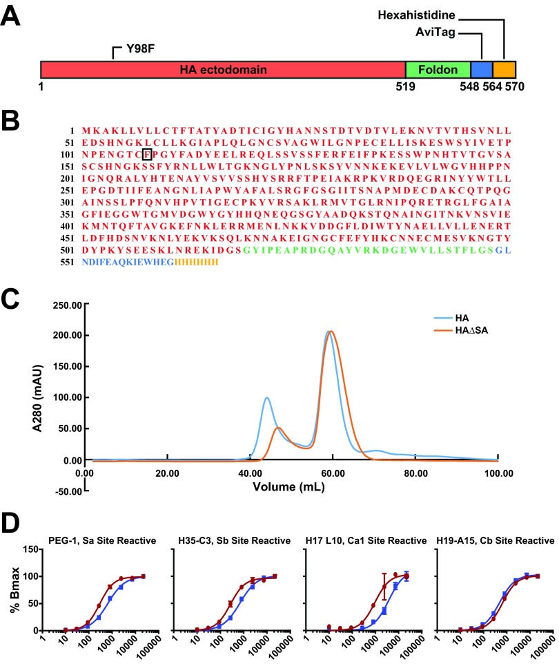
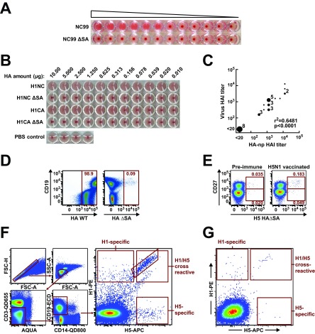
An understanding of the antigen-specific B-cell response to the influenza virus hemagglutinin (HA) is critical to the development of universal influenza vaccines, but it has not been possible to examine these cells directly because HA binds to sialic acid (SA) on most cell types. Here, we use structure-based modification of HA to isolate HA-specific B cells by flow cytometry and characterize the features of HA stem antibodies (Abs) required for their development. Incorporation of a previously described mutation (Y98F) to the receptor binding site (RBS) causes HA to bind only those B cells that express HA-specific Abs, but it does not bind nonspecifically to B cells, and this mutation has no effect on the binding of broadly neutralizing Abs to the RBS. To test the specificity of the Y98F mutation, we first demonstrated that previously described HA nanoparticles mediate hemagglutination and then determined that the Y98F mutation eliminates this activity. Cloning of immunoglobulin genes from HA-specific B cells isolated from a single human subject demonstrates that vaccination with H5N1 influenza virus can elicit B cells expressing stem monoclonal Abs (MAbs). Although these MAbs originated mostly from the IGHV1-69 germ line, a reasonable proportion derived from other genes. Analysis of stem Abs provides insight into the maturation pathways of IGVH1-69-derived stem Abs. Furthermore, this analysis shows that multiple non-IGHV1-69 stem Abs with a similar neutralizing breadth develop after vaccination in humans, suggesting that the HA stem response can be elicited in individuals with non-stem-reactive IGHV1-69 alleles.

Importance: Universal influenza vaccines would improve immune protection against infection and facilitate vaccine manufacturing and distribution. Flu vaccines stimulate B cells in the blood to produce antibodies that neutralize the virus. These antibodies target a protein on the surface of the virus called HA. Flu vaccines must be reformulated annually, because these antibodies are mostly specific to the viral strains used in the vaccine. But humans can produce broadly neutralizing antibodies. We sought to isolate B cells whose genes encode influenza virus antibodies from a patient vaccinated for avian influenza. To do so, we modified HA so it would bind only the desired cells. Sequencing the antibody genes of cells marked by this probe proved that the patient produced broadly neutralizing antibodies in response to the vaccine. Many sequences obtained had not been observed before. There are more ways to generate broadly neutralizing antibodies for influenza virus than previously thought.


Figures

FIG 1
FIG 1
Design and characterization of influenza virus HA-specific flow cytometry probe HAΔSA. (A) Crystal structures of HA in complex with SA analog or CH65 antibody illustrating interaction with tyrosine 98. Influenza virus HA (gray) is shown with one unit of the trimer colored (blue). The SA analog LSTc is shown bound to each unit. The circled area is shown enlarged, in complex with either SA (middle, green) or the CDR-H3 loop of the antibody CH65 (right, red). Tyrosine 98 makes polar contacts (dashed lines) with the adjacent histidine 183 and with SA (middle), but not the antibody (right). Mutation of tyrosine 98 to phenylalanine (right) removes the possibility of forming this polar contact with SA but has no effect on the interaction with the antibody. As the interaction with SA is weak (mM binding constant), removal of this polar contact eliminates binding. (B) HAΔSA mutation eliminates nonspecific binding. Wild-type (HA WT) or mutant (HA ΔSA) HA probes were conjugated to streptavidin-PE and used to label 293F cells grown in suspension. The no-probe control is shown in gray. Wild-type HA labels cells nonspecifically (top); HAΔSA does not (bottom). (C) Validation of the HAΔSA probe using cultured 293F cells transfected with membrane-bound IgM antibody genes. Cells transfected with the membrane-bound IgM form of HIV Env-specific control antibody VRC01, or RBS-directed antibody HA-specific CH65, or stem-directed antibody CR6261 express the respective antibody on the cell surface (anti-light-chain labeling, blue). Cells transfected with HA-directed antibody are labeled by HAΔSA, but those that express the anti-HIV antibody are not (HAΔSA labeling, red). The no-probe controls are shown in gray. (D) HAΔRBS labels only the stem-directed antibody CR6261, not the RBS-directed antibody CH65. The experiment was performed as described for that in panel C.
FIG 2
FIG 2
Schematic diagram and characterization of the HAΔSA probe. (A) Schematic diagram for HAΔSA probe. The ectodomain of HA (red), including the native N-terminal signal peptide, was fused to the T4 fibritin FoldOn (green), an AviTag (blue), and a hexahistidine purification tag (yellow). Numbering is according to the protein sequence of the construct. Position 98 of HA1 (HA numbering) was mutated to phenylalanine as described in the text. (B) Protein sequence for the H1/1999 HAΔSA probe, colored as in panel A. (C) Gel filtration chromatography of HA or HAΔSA on Superdex 200 16/60 column. HA and HAΔSA have similar elution profiles. Both elute at 58 ml, consistent with formation of the desired trimeric complex. A280, absorption at 280 nm; mAU, milliabsorbance units. (D) A panel of antibodies specific to the five antigenic sites of PR8 HA was tested against the PR8 HAΔSA probe and whole PR8 virus by ELISA titration. Representative ELISA curve plots for Sa, Sb, Ca1/Ca2, and Cb site-reactive antibodies are shown. Of the 25 tested, only one antibody, H37-76, was poorly reactive to the HAΔSA probe.
FIG 3
FIG 3
H1 and H5 HAΔSA probes identify B cells expressing strain-specific and cross-reactive antibodies against influenza virus HA. (A) Hemagglutination by lentivirus pseudotyped with H1N1 wild-type HA (NC99) or HAΔSA (NC99 ΔSA). Pseudovirus was diluted 1:4 from left to right. (B) Hemagglutination by HA-ferritin nanoparticles displaying the respective seasonal (NC99) or pandemic (CA09) H1N1 HA or HAΔSA. Pseudotyped lentivirus or HA-ferritin nanoparticles (np) (22) agglutinate red blood cells; the respective Y98F mutants of HA do not. Concentrations were determined by UV absorbance and serial dilutions performed from left to right as indicated. Formation of a dark red dot indicates failure to agglutinate at a given concentration. (C) Correlation between HAI measured with virus or HA np. Sera from ferrets vaccinated against A/New Caledonia/20/99 (H1N1) were tested against virus or strain-matched HA np. (D) Validation of the HAΔSA probe using B cells from a human donor. Human donor PBMCs were analyzed by flow cytometry. The wild-type probe labels nearly all cells (left); the HAΔSA probe does not (right). Probes were purified and labeled in the same manner, and identical concentrations of each probe were used to label the cells. (E) Effect of vaccination for H5N1 influenza virus on the frequency of H5+ cells. PBMC samples from one subject (VRC 310-036) drawn before vaccination for H5N1 (left), or 2 weeks afterward (right), were labeled with the HAΔSA probe corresponding to the vaccine strain, the B-cell maturation marker CD27, and other B-cell markers as described in Materials and Methods. Vaccination against H5N1 influenza virus increases the proportion of antigen-specific CD19+ CD27 and CD19+ CD27+ B cells, detected by the H5 HAΔSA probe. (F) Sorting for H1/H5 cross-reactive B cells. PMBCs were sequentially gated to isolate lymphocytes and to remove doublets, CD3+ T cells, CD14+ monocytes, and dead cells stained by the Aqua viability dye. Antigen specificity of CD19+ B cells was assessed using binding to H1 HAΔSA conjugated to streptavidin-PE and H5 HAΔSA conjugated to streptavidin-APC. A set of 92 single B cells from a single subject (VRC 310-018) were index-sorted and subjected to antibody gene sequencing. (G) PBMCs from a prevaccination sample from the same patient were analyzed as in panel F.
FIG 4
FIG 4
H1/H5 cross-reactive antibodies bind H1 and H5 HAs and neutralize HA-pseudotyped lentivirus vectors. (A) Monoclonal antibodies for 22 selected sequences were expressed and tested by ELISA for binding to H1 or H5 HA, for HAI, and for their ability to neutralize replication-defective lentiviral vector pseudotyped with HA from the various H1 and H5 strains indicated. Binding is expressed as the base-10 logarithm of the concentration in μg/ml. HAI is expressed as the minimum concentration at which agglutination is inhibited in μg/ml; none of the cloned antibodies showed HAI activity. Neutralization is reported as the 50% inhibitory concentration (IC50) in μg/ml. Most antibodies neutralize all tested strains similarly. For unknown reasons, the pseudovirus made from the H5N1 A/Indonesia/05/2005 strain used for vaccination in this study is not neutralized well by the antibodies tested, nor by the control antibody CR6261. 2D1 and 9E8 are head-specific monoclonal antibodies against H1 and H5, respectively (28, 36). Clonally related antibodies are indicated. (B) Genetic characteristics of further H1/H5 cross-reactive antibodies not expressed. id, identifier; nd, tests not performed; VK, variable kappa-chain gene; Vγ, variable lambda-chain gene.
FIG 5
FIG 5
Sequences of novel IGHV1-69 cross-reactive antibodies are consistent with the structural basis for development of broadly neutralizing influenza virus antibodies. (A) Multiple sequence alignment of cloned IGHV1-69 VH gene segments compared to germ line alleles present in this subject, 1-69*01 and -*06 (green) and CR6261 (somatically mutated residues in blue). Residues conserved from the IGHV1-69-encoded protein are denoted by dots; somatically mutated residues in each antibody are denoted by black letters or, when congruent with CR6261, by blue letters. Shaded areas correspond to colors in panel B. (B) Structure of the HA stem epitope (gray surface) in complex with the VH portion of CR6261 (contact residues shown as colored sticks). The CDR-H3 also contributes two non-germ line-encoded contacts (not shown). Germ line-encoded residues are drawn in green; somatically mutated residues in blue. Pie charts represent the observed frequency of each amino acid at the residue indicated; the color of the center of each pie chart corresponds to the shading used in the sequence alignment in panel A; the wedges of each pie chart are colored green to denote the germ line residue, or blue to denote the residue found in CR6261, or white if other. The previously described structural basis for development of this class of antibody included strict conservation of F29 and F54, preference for hydrophobicity at F53, mutations at 28 and 30 that stabilize the bound conformation of the CDR-H1 (arginine [R] and the next most frequent residue, asparagine [N], are chemically similar) and, optionally, mutation of residue 74 to phenylalanine [F].

Similar articles

Cited by

References

    1. Ekiert DC, Bhabha G, Elsliger MA, Friesen RH, Jongeneelen M, Throsby M, Goudsmit J, Wilson IA. 2009. Antibody recognition of a highly conserved influenza virus epitope. Science 324:246–251. 10.1126/science.1171491 - DOI - PMC - PubMed
    1. Sui J, Hwang WC, Perez S, Wei G, Aird D, Chen LM, Santelli E, Stec B, Cadwell G, Ali M, Wan H, Murakami A, Yammanuru A, Han T, Cox NJ, Bankston LA, Donis RO, Liddington RC, Marasco WA. 2009. Structural and functional bases for broad-spectrum neutralization of avian and human influenza A viruses. Nat. Struct. Mol. Biol. 16:265–273. 10.1038/nsmb.1566 - DOI - PMC - PubMed
    1. Corti D, Voss J, Gamblin SJ, Codoni G, Macagno A, Jarrossay D, Vachieri SG, Pinna D, Minola A, Vanzetta F, Silacci C, Fernandez-Rodriguez BM, Agatic G, Bianchi S, Giacchetto-Sasselli I, Calder L, Sallusto F, Collins P, Haire LF, Temperton N, Langedijk JP, Skehel JJ, Lanzavecchia A. 2011. A neutralizing antibody selected from plasma cells that binds to group 1 and group 2 influenza A hemagglutinins. Science 333:850–856. 10.1126/science.1205669 - DOI - PubMed
    1. Ekiert DC, Friesen RH, Bhabha G, Kwaks T, Jongeneelen M, Yu W, Ophorst C, Cox F, Korse HJ, Brandenburg B, Vogels R, Brakenhoff JP, Kompier R, Koldijk MH, Cornelissen LA, Poon LL, Peiris M, Koudstaal W, Wilson IA, Goudsmit J. 2011. A highly conserved neutralizing epitope on group 2 influenza A viruses. Science 333:843–850. 10.1126/science.1204839 - DOI - PMC - PubMed
    1. Whittle JR, Zhang R, Khurana S, King LR, Manischewitz J, Golding H, Dormitzer PR, Haynes BF, Walter EB, Moody MA, Kepler TB, Liao HX, Harrison SC. 2011. Broadly neutralizing human antibody that recognizes the receptor-binding pocket of influenza virus hemagglutinin. Proc. Natl. Acad. Sci. U. S. A. 108:14216–14221. 10.1073/pnas.1111497108 - DOI - PMC - PubMed

Publication types

MeSH terms

Associated data