

Increased stability and limited proliferation of CD4+ central memory T cells differentiate nonprogressive simian immunodeficiency virus (SIV) infection of sooty mangabeys from progressive SIV infection of rhesus macaques
- PMID: 24501416
- PMCID: PMC3993768
- DOI: 10.1128/JVI.03515-13
Increased stability and limited proliferation of CD4+ central memory T cells differentiate nonprogressive simian immunodeficiency virus (SIV) infection of sooty mangabeys from progressive SIV infection of rhesus macaques
Abstract
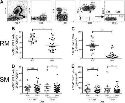
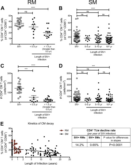
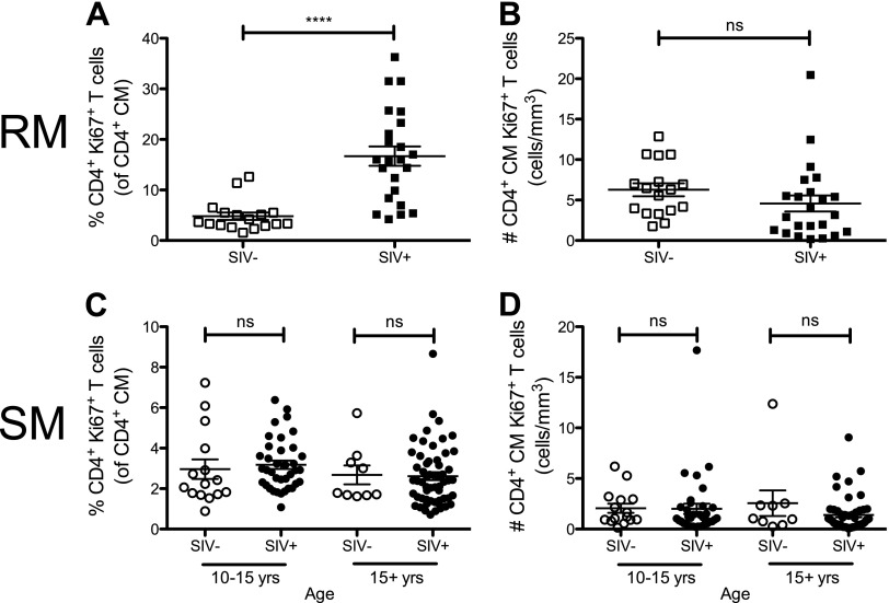
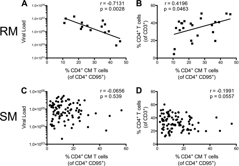
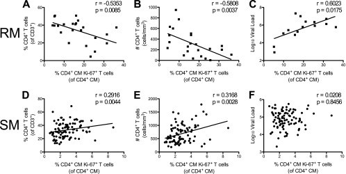
Depletion of CD4(+) central memory T (TCM) cells dictates the tempo of progression to AIDS in simian immunodeficiency virus (SIV)-infected rhesus macaques (RMs) both in the natural history of infection and in the context of vaccination. CD4(+) TCM cells of sooty mangabeys (SMs), a natural host for SIV in which infection is nonpathogenic, are less susceptible to SIV infection than CD4(+) TCM cells of RMs. Whether this relative protection from infection translates into increased stability of CD4(+) TCM cells in natural versus nonnatural hosts has not yet been determined. Here we compared, both cross-sectionally and longitudinally, the levels of CD4(+) TCM cells in a large cohort of SMs and RMs and the association between CD4(+) TCM levels and the main virologic and immunologic markers of disease progression. Consistent with their lower susceptibility to infection, CD4(+) TCM cells of SIV-infected SMs are lost with kinetics 20 times slower than those of SIV-infected RMs. Remarkably, the estimated length of time of SIV infection needed for CD4(+) TCM cells to fall to half of their initial levels is <16 months for RMs but >17 years for SMs. Furthermore, the fraction of proliferating CD4(+) TCM cells is significantly lower in SIV-infected SMs than in SIV-infected RMs, and the extent of CD4(+) TCM cell proliferation is associated positively with CD4(+) T cell levels in SIV-infected SMs but negatively with CD4(+) T cell levels in SIV-infected RMs. Collectively, these findings identify increased stability and maintenance of the prohomeostatic role of CD4(+) TCM cells as features distinguishing nonprogressive from progressive SIV infections and support the hypothesis of a direct mechanistic link between the loss of CD4(+) TCM cells and disease progression.
Importance: Comparison of the immunologic effects of simian immunodeficiency virus (SIV) infection on rhesus macaques (RMs), a species characterized by progression to AIDS, and natural host sooty mangabeys (SMs), a species which remains AIDS free, has become a useful tool for identifying mechanisms of human immunodeficiency virus (HIV) disease progression. One such distinguishing feature is that CD4(+) central memory T (TCM) cells in SIV-infected SMs are less infected than the same cells in RMs. Here we investigated whether lower levels of infection in SMs translate into a better-preserved CD4(+) TCM compartment. We found that the CD4(+) TCM compartment is significantly more stable in SIV-infected SMs. Likely to compensate for this cell loss, we also found that CD4(+) TCM cells increase their level of proliferation upon SIV infection in RMs but not in SMs, which mechanistically supports their preferential infectivity. Our study provides new insights into the importance of long-term maintenance of CD4(+) TCM homeostasis during HIV/SIV infection.
Figures
Similar articles
-
Reduced Simian Immunodeficiency Virus Replication in Macrophages of Sooty Mangabeys Is Associated with Increased Expression of Host Restriction Factors.J Virol. 2015 Oct;89(20):10136-44. doi: 10.1128/JVI.00710-15. Epub 2015 Jul 22. J Virol. 2015. PMID: 26202248 Free PMC article.
-
Depletion of CD8+ cells in sooty mangabey monkeys naturally infected with simian immunodeficiency virus reveals limited role for immune control of virus replication in a natural host species.J Immunol. 2007 Jun 15;178(12):8002-12. doi: 10.4049/jimmunol.178.12.8002. J Immunol. 2007. PMID: 17548637
-
Antiretroviral Therapy in Simian Immunodeficiency Virus-Infected Sooty Mangabeys: Implications for AIDS Pathogenesis.J Virol. 2016 Jul 27;90(16):7541-7551. doi: 10.1128/JVI.00598-16. Print 2016 Aug 15. J Virol. 2016. PMID: 27279614 Free PMC article.
-
Naturally SIV-infected sooty mangabeys: are we closer to understanding why they do not develop AIDS?J Med Primatol. 2005 Oct;34(5-6):243-52. doi: 10.1111/j.1600-0684.2005.00122.x. J Med Primatol. 2005. PMID: 16128919 Review.
-
What can natural infection of African monkeys with simian immunodeficiency virus tell us about the pathogenesis of AIDS?AIDS Rev. 2004 Jan-Mar;6(1):40-53. AIDS Rev. 2004. PMID: 15168740 Review.
Cited by
-
Plasma Levels of C-Type Lectin REG3α and Gut Damage in People With Human Immunodeficiency Virus.J Infect Dis. 2020 Jan 1;221(1):110-121. doi: 10.1093/infdis/jiz423. J Infect Dis. 2020. PMID: 31504638 Free PMC article.
-
Proteome and Protein Network Analyses of Memory T Cells Find Altered Translation and Cell Stress Signaling in Treated Human Immunodeficiency Virus Patients Exhibiting Poor CD4 Recovery.Open Forum Infect Dis. 2016 Mar 15;3(2):ofw037. doi: 10.1093/ofid/ofw037. eCollection 2016 Mar. Open Forum Infect Dis. 2016. PMID: 28293663 Free PMC article.
-
Viremic non-progression in HIV/SIV infection: A tied game between virus and host.Cell Rep Med. 2025 Jan 21;6(1):101921. doi: 10.1016/j.xcrm.2024.101921. Cell Rep Med. 2025. PMID: 39842407 Free PMC article. Review.
-
Limited HIV infection of central memory and stem cell memory CD4+ T cells is associated with lack of progression in viremic individuals.PLoS Pathog. 2014 Aug 28;10(8):e1004345. doi: 10.1371/journal.ppat.1004345. eCollection 2014 Aug. PLoS Pathog. 2014. PMID: 25167059 Free PMC article.
-
Nonprogressing HIV-infected children share fundamental immunological features of nonpathogenic SIV infection.Sci Transl Med. 2016 Sep 28;8(358):358ra125. doi: 10.1126/scitranslmed.aag1048. Sci Transl Med. 2016. PMID: 27683550 Free PMC article.
References
-
- Dunham R, Pagliardini P, Gordon S, Sumpter B, Engram J, Moanna A, Paiardini M, Mandl JN, Lawson B, Garg S, McClure HM, Xu YX, Ibegbu C, Easley K, Katz N, Pandrea I, Apetrei C, Sodora DL, Staprans SI, Feinberg MB, Silvestri G. 2006. The AIDS resistance of naturally SIV-infected sooty mangabeys is independent of cellular immunity to the virus. Blood 108:209–217. 10.1182/blood-2005-12-4897 - DOI - PMC - PubMed
Publication types
MeSH terms
Grants and funding
LinkOut - more resources
Full Text Sources
Other Literature Sources
Research Materials
