. 2014 Apr;25(7):1073-96.
doi: 10.1091/mbc.E13-09-0521. Epub 2014 Feb 5.

Caenorhabditis elegans HOPS and CCZ-1 mediate trafficking to lysosome-related organelles independently of RAB-7 and SAND-1

Affiliations

Caenorhabditis elegans HOPS and CCZ-1 mediate trafficking to lysosome-related organelles independently of RAB-7 and SAND-1

Jared L Delahaye et al. Mol Biol Cell. 2014 Apr.

Abstract

As early endosomes mature, the SAND-1/CCZ-1 complex acts as a guanine nucleotide exchange factor (GEF) for RAB-7 to promote the activity of its effector, HOPS, which facilitates late endosome-lysosome fusion and the consumption of AP-3-containing vesicles. We show that CCZ-1 and the HOPS complex are essential for the biogenesis of gut granules, cell type-specific, lysosome-related organelles (LROs) that coexist with conventional lysosomes in Caenorhabditis elegans intestinal cells. The HOPS subunit VPS-18 promotes the trafficking of gut granule proteins away from lysosomes and functions downstream of or in parallel to the AP-3 adaptor. CCZ-1 also acts independently of AP-3, and ccz-1 mutants mistraffic gut granule proteins. Our results indicate that SAND-1 does not participate in the formation of gut granules. In the absence of RAB-7 activity, gut granules are generated; however, their size and protein composition are subtly altered. These observations suggest that CCZ-1 acts in partnership with a protein other than SAND-1 as a GEF for an alternate Rab to promote gut granule biogenesis. Point mutations in GLO-1, a Rab32/38-related protein, predicted to increase spontaneous guanine nucleotide exchange, specifically suppress the loss of gut granules by ccz-1 and glo-3 mutants. GLO-3 is known to be required for gut granule formation and has homology to SAND-1/Mon1-related proteins, suggesting that CCZ-1 functions with GLO-3 upstream of the GLO-1 Rab, possibly as a GLO-1 GEF. These results support LRO formation occurring via processes similar to conventional lysosome biogenesis, albeit with key molecular differences.


Figures
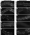
FIGURE 1:
FIGURE 1:
Autofluorescent compartments in adult intestinal cells. The 9-μm maximum-intensity projections, which span 50% the width of the intestine, of GFP channel autofluorescence centered on the lumen of the anterior intestine show that, in comparison to (A) wild type, the (B) vps-11, (C) vps-16, (E) vps-33, (F) vps-39, (G) vps-41, and (I) ccz-1 mutants exhibited reduced numbers of autofluorescent compartments. Mutations in (D) vps-18, (H) sand-1, and (J) rab-7 did not obviously alter the number of autofluorescent organelles. Black arrows denote representative autofluorescent compartments.
FIGURE 2:
FIGURE 2:
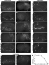
Analysis of birefringent and acidified gut granules in embryonic intestinal cells. Embryos were analyzed with polarization microscopy to detect birefringent material, stained with LysoSensor Green, and analyzed with fluorescence microscopy to detect acidified compartments. (A, F) Wild-type, (C, H) sand-1, and (D, I) rab-7 mutant embryos displayed birefringent and acidified gut granules (white arrows). (B, G) ccz-1 and (E, J) vps-18 mutants lacked acidified and birefringent gut granules and mislocalized birefringent material into the intestinal lumen (black arrows). (K–O) RNAi targeting subunits of the VPS-C/HOPS complex in an rrf-3(pk1426) RNAi–sensitive strain caused a reduction in the number of birefringent organelles (white arrows) and sometimes led to misaccumulation of birefringent material in the intestinal lumen (black arrows). (P, Q) Birefringent material (white arrows) was present within the intestinal cells of vps-18 mutants at 15°C and was distributed throughout the embryo at 25°C. The intestine is flanked by black arrowheads in A–P. Pretzel-stage embryos are shown in A–C, E, and K–P; 1.5-fold-stage embryos are shown in D and F–J. A terminally arrested pre–bean stage vps-18 mutant embryo is shown in Q. Embryos are ∼50 μm in length. (R) Individual vps-18 mutant embryos were shifted from 15 to 22°C at the indicated stage, and on reaching threefold stage or later, were analyzed using polarization microscopy. Embryos exhibiting <20 birefringent granules were scored as having a reduced number of granules. E2, E4, and E8 refer to the number of intestinal cells in the intestinal primordium; early-bean-stage embryos are E16 before the apical migration of intestinal nuclei. Bean and 1.5-fold stages refer to the body morphology. The number of embryos scored is indicated in parentheses near each data point. Unshifted vps-18 mutants grown at 15°C exhibited a Glo phenotype only 9% of the time (n = 33). Wild-type embryos that underwent similar temperature shifts always displayed >50 birefringent granules.
FIGURE 3:
FIGURE 3:
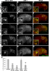
The distribution of CDF-2::GFP and PGP-2 is altered in vps-18 and ccz-1 mutants. Anti–PGP-2 antibodies and ectopically expressed CDF-2::GFP colocalized at gut granules in (A–C) wild type and (J–L) sand-1 and (M-O) rab-7 mutants (black arrows within insets). (D–F) CDF-2::GFP–containing organelles in vps-18 mutants lacked PGP-2 staining (white arrows in insets). (G–I) The majority of CDF-2::GFP–labeled compartments in ccz-1 mutants lacked anti–PGP-2 staining (white arrows in insets). In A–O, 1.5-fold-stage embryos are shown, black arrowheads flank the intestine, and the insets are 5 μm wide. (P) For each genotype, at least 25 randomly selected CDF-2::GFP–containing intestinal compartments in five different 1.5-fold-stage embryos were scored for the presence of anti-PGP-2 signals. The mean is plotted, and error bars represent the 95% confidence limit. A one-way analysis of variance (ANOVA) comparing each mutant to wild type was used to calculate p values (**p ≤ 0.001).
FIGURE 4:
FIGURE 4:
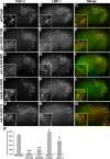
The distribution of PGP-2 and LMP-1 is altered in vps-18, ccz-1, and rab-7 mutants. Antibodies recognizing PGP-2 and LMP-1 colocalized at gut granules in (A–C) wild type and (J–L) sand-1 mutants (black arrows in insets). (D–I) PGP-2–stained organelles in vps-18 and ccz-1 mutants lacked LMP-1 staining (white arrows within insets). (M–O) In rab-7 mutants, LMP-1 staining was weakly present on some (black arrows in insets) but not all (white arrows in insets) anti-PGP-2–marked gut granules. In A–O, 1.5-fold-stage embryos are shown, black arrowheads flank the intestine, and insets are 5 μm wide. (P) For each genotype, at least 25 randomly selected PGP-2–stained intestinal compartments in five different 1.5-fold-stage embryos were scored for the presence of LMP-1 staining. The mean is plotted, and error bars represent the 95% confidence limit. A one-way ANOVA comparing each mutant to wild type was used to calculate p values (*p ≤ 0.05, **p ≤ 0.001).
FIGURE 5:
FIGURE 5:
The distribution of CDF-2::GFP and LMP-1 is altered in vps-18, ccz-1, and rab-7 mutants. Nearly all CDF-2::GFP–containing organelles were marked by anti–LMP-1 antibodies in (A–C) wild-type and (J–L) sand-1 mutant cells (black arrows in insets). In (D–F) vps-18, (G–I) ccz-1, and (M–O) rab-7 mutants, a subset of CDF-2::GFP–marked organelles contained anti–LMP-1 staining (black arrows in insets), although most lacked LMP-1 (white arrows in insets). In A–L, 1.5-fold-stage embryos are shown, and in M–O, a late-bean-stage embryo is shown. In A–O, black arrowheads flank the intestine and insets are 5 μm wide. (P) For each genotype, at least 25 randomly selected CDF-2::GFP–containing intestinal compartments in five different 1.5-fold-stage embryos were scored for the presence of LMP-1 staining. The mean is plotted, and error bars represent the 95% confidence limit. A one-way ANOVA comparing each mutant to wild type was used to calculate p values (*p ≤ 0.05, **p ≤ 0.001).
FIGURE 6:
FIGURE 6:
CDF-2::GFP is mislocalized to endolysosomes in vps-18 mutants. (A–C, G–I) Endogenous RAB-7 detected with anti–RAB-7 antibodies was not associated with CDF-2::GFP–containing organelles in wild type or ccz-1 mutants (white arrows in insets). (D–F) In vps-18 mutants, anti–RAB-7 staining was often localized to CDF-2::GFP–marked compartments (black arrows in insets). The lysosomal hydrolase F11E6.1a::mCherry did not appreciably localize to CDF-2::GFP–marked compartments in (J–L) wild type or (P–R) ccz-1 mutants (white arrows in insets). (M–O) In vps-18 mutants, F11E6.1a::mCherry often colocalized with CDF-2::GFP–containing compartments (black arrows in insets). In A–R, single optical sections of 1.5-fold-stage embryos are shown, black arrowheads flank the intestine, and insets are 5 μm wide. In A–F, white arrowheads denote the apical surface of intestinal cells. (S, T) For each genotype, at least 25 randomly selected CDF-2::GFP–containing intestinal compartments in five different 1.5-fold-stage embryos were scored for the presence of RAB-7 or F11E6.1a::mCherry. The mean is plotted, and error bars represent the 95% confidence limit. A one-way ANOVA comparing each mutant to wild type was used to calculate p values (*p ≤ 0.05, **p ≤ 0.001).
FIGURE 7:
FIGURE 7:
Analysis of LMP-1::GFP trafficking. (A–E) Mutations in (B) vps-18, (C) ccz-1, and to a lesser extent (E) rab-7 led to an increased number of LMP-1::GFP–marked organelles. (R) The total number of LMP-1::GFP compartments was quantified in the four cells that compose Int2 and Int3 (marked by black arrows in A–E) in five different 1.5-fold-stage embryos of each genotype. The mean is plotted, and error bars represent the 95% confidence limit. A one-way ANOVA comparing each mutant to wild type was used to calculate p values (*p ≤ 0.05, **p ≤ 0.001). (F–H) Relative to wild type, a higher proportion of LMP-1::GFP compartments in (I–K) vps-18 mutants contained RAB-7 antibody signals, and a decreased proportion of LMP-1::GFP compartments in (L–N) ccz-1 and sand-1 mutants contained RAB-7 (in the insets, black arrows denote LMP-1::GFP compartments marked by RAB-7 antibodies and white arrows label compartments lacking a RAB-7 signal). (S) For each genotype, at least 25 randomly selected LMP-1::GFP–containing intestinal compartments in five different 1.5-fold-stage embryos were scored for the presence of signal from the RAB-7 antibody. The mean is plotted, and error bars represent the 95% confidence limit. A one-way ANOVA comparing each mutant to wild type was used to calculate p (*p ≤ 0.05, **p ≤ 0.001). In A–E, 1-μm maximum-intensity projections, and in F–Q, single optical sections, of 1.5-fold-stage embryos are shown. The white arrowhead in B denotes the apical cell membrane. In images showing embryos, the black arrowheads flank the intestine. The insets are 5 μm wide.
FIGURE 8:
FIGURE 8:
Effects of disrupting vps-18 and ccz-1 function on gut granule protein trafficking in apt-7 mutants. Embryos expressing CDF-2::GFP were stained with anti–PGP-2 antibodies. (A–C) In apt-7 single mutants, CDF-2::GFP–labeled compartments were rarely marked by PGP-2 antibodies (white and black arrows in insets). (D–I) Relative to the single mutants, apt-7; vps-18 and apt-7; ccz-1 double mutants displayed an increased proportion of organelles that contained both CDF-2::GFP and PGP-2 (black arrows in insets). (J) For each genotype, at least 25 randomly selected CDF-2::GFP–containing intestinal compartments in five different 1.5-fold-stage embryos were scored for the presence of PGP-2 staining. The mean is plotted, and error bars represent the 95% confidence limit. A one-way ANOVA comparing the indicated genotypes was used to calculate p values (*p ≤ 0.05, **p ≤ 0.001). In A–I, 1.5-fold-stage embryos are shown, and black arrowheads flank the intestine. The insets are 5 μm wide.
FIGURE 9:
FIGURE 9:
Expression of GFP::GLO-1(ΔG4) mutants leads to increased numbers of autofluorescent and acidified gut granules in ccz-1 and glo-3 mutants. Young adults were analyzed for the presence of autofluorescent material or the accumulation of the acidophilic dye acridine orange. (A, J) Wild type contained many autofluorescent and acridine orange–stained organelles. (B, C, F, G, K, L, O, and P) ccz-1 and glo-3 mutants contained few autofluorescent and acridine orange–stained gut granules, whose numbers did not increase upon the expression of gfp::glo-1 under control of the vha-6 promoter. (D, E, H, I, M, N, Q, and R) The addition of glo-1(D132A) or glo-1(I133F), which contain mutations in the G4 motif of GLO-1, led to increased numbers of autofluorescent and acridine orange–labeled compartments in ccz-1 and glo-3 mutants. Maximum-intensity projections of fluorescence signals spanning the entire depth of the anterior intestine are shown. White arrows denote autofluorescent or acridine orange–stained organelles. (S) Alignment of the G4 domain of C. elegans (Ce) GLO-1 and related Rab proteins from humans (Hs) and yeast (Sc), denoting the location of mutations predicted to alter guanine nucleotide binding. (T) Alignment of the amino-terminal region of C. elegans GLO-3 (Ce) and HPS-1 from D. melanogaster (Dm) and humans (Hs). Location of β-sheets and α-helices that compose the Longing domain in HPS1 proteins are present below the sequences. A consensus of GLO-3 secondary structural predictions for β-sheets (listed as E) and α-helices (listed as H) are positioned above the sequences.

Similar articles

Cited by

References

    1. Akbar MA, Ray S, Kramer H. The SM protein Car/Vps33A regulates SNARE-mediated trafficking to lysosomes and lysosome-related organelles. Mol Biol Cell. 2009;20:1705–1714. - PMC - PubMed
    1. Altschul SF, Madden TL, Schaffer AA, Zhang J, Zhang Z, Miller W, Lipman DJ. Gapped BLAST and PSI-BLAST: a new generation of protein database search programs. Nucleic Acids Res. 1997;25:3389–3402. - PMC - PubMed
    1. Ambrosio AL, Boyle JA, Di Pietro SM. Mechanism of platelet dense granule biogenesis: study of cargo transport and function of Rab32 and Rab38 in a model system. Blood. 2012;120:4072–4081. - PMC - PubMed
    1. Angers CG, Merz AJ. HOPS interacts with Apl5 at the vacuole membrane and is required for consumption of AP-3 transport vesicles. Mol Biol Cell. 2009;20:4563–4574. - PMC - PubMed
    1. Aoyama M, Sun-Wada GH, Yamamoto A, Yamamoto M, Hamada H, Wada Y. Spatial restriction of bone morphogenetic protein signaling in mouse gastrula through the mVam2-dependent endocytic pathway. Dev Cell. 2012;22:1163–1175. - PubMed

Publication types

MeSH terms

LinkOut - more resources