. 2014 Feb 27;57(4):1289-98.
doi: 10.1021/jm401471v. Epub 2014 Feb 6.

Optimization of the lactam side chain of 7-azaindenoisoquinoline topoisomerase I inhibitors and mechanism of action studies in cancer cells

Affiliations

Optimization of the lactam side chain of 7-azaindenoisoquinoline topoisomerase I inhibitors and mechanism of action studies in cancer cells

Evgeny Kiselev et al. J Med Chem. .

Abstract

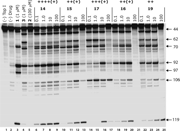
Optimization of the lactam ω-aminoalkyl substituents in a series of 7-azaindenoisoquinolines resulted in new anticancer agents with improved Top1 inhibitory potencies and cancer cell cytotoxicities. The new compounds 14-17 and 19 exhibited mean graph midpoint cytotoxicity (GI50) values of 21-71 nM in the NCI panel of 60 human cancer cell cultures. Ternary 7-azaindenoisoquinoline-DNA-Top1 cleavage complexes that persist for up to 6 h were detected in HCT116 colon cancer cells. Ternary complexes containing 7-azaindenoisoquinolines were significantly more stable than those in which camptothecin was incorporated. DNA content distribution histograms showed S-phase block 3 h after drug removal. Drug-induced DNA damage in HCT116 cells was revealed by induction of the histone γ-H2AX marker. The 7-azaindenoisoquinolines were able to partially overcome resistance in several drug-resistant cell lines, and they were not substrates for the ABCB1 drug efflux transporter. Molecular modeling studies indicate that the 7-azaindenoisoquinolines intercalate at the DNA cleavage site in DNA-Top1 covalent complexes with the lactam side chain projecting into the major groove. Overall, the results indicate that the 7-azaindenoisoquinolines are promising anticancer agents that merit further development.


Figures

Figure 1
Figure 1
Representative Top1 inhibitors.
Scheme 1
Scheme 1
Scheme 2
Scheme 2
Figure 2
Figure 2
Lane 1: DNA alone. Lane 2: Top1 alone. Lane 3: Top1 and 1 (1 μM). Lane 4: Top1 and 3 (1 μM). Lane 5: Top1 and 2 (100 μM). Lanes 6–9: Top1 and 14 at 0.1, 1, 10, and 100 μM, respectively. Lanes 10–13: Top1 and 15 at 0.1, 1, 10, and 100 μM, respectively. Lanes 14–17: Top1 and 17 at 0.1, 1, 10, and 100 μM, respectively. Lanes 18–21: Top1 and 16 at 0.1, 1, 10, and 100 μM, respectively. Lanes 22–25: Top1 and 19 at 0.1, 1, 10, and 100 μM, respectively. The numbers and arrows on the right show the cleavage site positions.
Figure 3
Figure 3
Structural formulas of topotecan and doxorubicin.
Figure 4
Figure 4
(A) Top1cc’s induced after treatment for 1 h with 11 (0.1, 1, and 10 μM, bottom row of the left panel) or 14 (0.1, 1, and 10 μM, bottom row of the right panel), 1 μM 1, and 1 μM 20. ND means no drug. (B) Persistent Top1cc’s produced by treatment for 1 h with 11 and 14 and assayed at the indicated time points after incubation in drug-free medium. (C) Representative alkaline elution experiments showing persistent Top1cc’s measured as DNA protein cross-links (DPCs) in cells treated with 11 or 14. DPCs persisting 1 h after drug removal are shown. Rapidly reversible DPCs induced by CPT were used as a control. Untreated cells receiving only 30 Gy of irradiation to induce strand breaks were used as a negative control. The fraction of DNA remaining on the filter is plotted vs time (hours).
Figure 5
Figure 5
Cell cycle analysis of HCT116 cells treated with 1 μM 1, 11, or 14 for 1 h (column 1) followed by incubation in drug-free medium for the indicated times (columns 2–6). Fixed cells were stained with propidium iodide (PI) and analyzed for DNA content distribution histograms by flow cytometry. The 48 h reversal time point is included in the inset to show cells collected from the supernatant.
Figure 6
Figure 6
DNA damage induced by nitro-azaindenoisoquinolines. Drug-induced DNA damage was measured by induction of histone γ-H2AX following treatment for 1 h with 1 μM 1, 11, or 14. γ-H2AX-positive cells induced by 1, 11, or 14 were analyzed by flow cytometry. Numbers above each profile represent the percent cells that score for γ-H2AX. The DNA content determined by propidium iodide (PI) is on the bottom axis.
Figure 7
Figure 7
Hypothetical mode of binding of 14 (green) to Top1cc (gray). Hydrogen bonds are presented as distances between corresponding heavy atoms. A DNA base pair was removed from the top to clarify the view. This figure was generated by molecular modeling starting from the X-ray crystal structure of an indenoisoquinoline–Top1–DNA complex obtained from the Protein Data Bank (entry 1SC7).

References

    1. Wall M. E.; Wani M. C.; Cook C. E.; Palmer K. H.; McPhail A. T.; Sim G. A. Plant Antitumor Agents. I. The Isolation and Structure of Camptothecin, a Novel Alkaloidal Leukemia and Tumor Inhibitor from Camptotheca acuminata. J. Am. Chem. Soc. 1966, 88, 3888–3890.
    1. Kohlhagen G.; Paull K.; Cushman M.; Nagafuji P.; Pommier Y. Protein-Linked DNA Strand Breaks Induced by NSC 314622, a Novel Noncamptothecin Topoisomerase I Poison. Mol. Pharmacol. 1998, 54, 50–58. - PubMed
    1. Khadka D. B.; Cho W.-J. Topoisomerase Inhibitors as Anticancer Agents: A Patent Update. Expert Opin. Ther. Pat. 2013, 23, 1033–1056. - PubMed
    1. Staker B. L.; Feese M. D.; Cushman M.; Pommier Y.; Zembower D.; Stewart L.; Burgin A. B. Structures of Three Classes of Anticancer Agents Bound to the Human Topoisomerase I-DNA Covalent Complex. J. Med. Chem. 2005, 48, 2336–2345. - PubMed
    1. Pommier Y.; Marchand C. Interfacial Inhibitors: Targeting Macromolecular Complexes. Nat. Rev. Drug Discovery 2012, 11, 25–36. - PMC - PubMed

Publication types

MeSH terms