

Review
doi: 10.1089/hum.2014.2501.
Herpes simplex viral vectors: late bloomers with big potential
Affiliations
- PMID: 24502405
- PMCID: PMC3922308
- DOI: 10.1089/hum.2014.2501
Item in Clipboard
Review
Herpes simplex viral vectors: late bloomers with big potential
Hum Gene Ther.
2014 Feb.
No abstract available
Figures
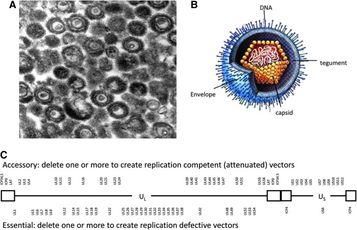
HSV-1 particle and genome structure. (A) Electron micrograph of purified HSV particles reveals the envelope, capsid, and condensed DNA. (B) Representation of the particle locates the envelope with spike glycoproteins, tegument, regular icosahedral capsid, and internal DNA (reproduced from the thesis of Paola Grandi, University of Ferrara, 2002). (C) The linear double-stranded DNA is shown as a diagram. Boxes represent the inverted terminal repeats that flank the unique long (UL) and unique short (US) sequences. Accessory viral genes (upper) can be manipulated to create attenuated viruses, and essential genes (lower) can be deleted to make replication-defective viruses. HSV-1, herpes simplex virus type 1.

Genome structure: location of gene classes. (A) Diagram of the linear viral genome. Blue boxes, inverted repeats. Terminal repeat (TR) and internal repeat (IR) flanking the long (L) and short (S) unique segments. (B) The viral transcription units are color coded as immediate early (IE, green), early (E, yellow), early-late (L1, red), late (L2, brown), and latency (LAT) gene (purple). The blue boxes represent the repeat regions that can be manipulated to construct replication-defective vectors. The internal joint region contains genes that are duplicated in the terminal repeats and can be deleted for insertion of foreign sequences. The IE genes are deleted to create completely silent vectors. The LAT region contains elements that can be used for insertion of transgenes that remain active in the absence of the IE genes.
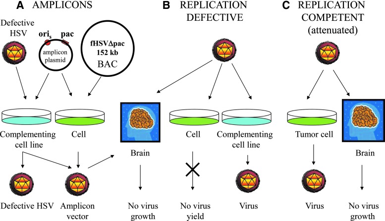
HSV vector design strategies. (A) Amplicon designs. Amplicons are genome-length plasmid-like vectors that contain a viral origin (ori) and packaging (pac) sequences that allow amplification of transfected amplicon DNA and incorporation into virus particles for amplicon delivery to cells. Replication-defective helper virus supplies viral functions needed to replicate amplicon DNA and to make helper and amplicon particles in complementing (blue) cells. Cotransfection of noncomplementing cells (green) with amplicon DNA and helper DNA lacking packaging sequences will produce helper-free amplicon particles. (B) Replication-defective vector designs. Replication-defective viruses are deleted for one or more essential virus genes and must be grown on cell lines that complement these deleted functions in trans (blue). Infection of noncomplementing cells in culture will not produce virus (green), and infection of other tissues (e.g., brain) will not allow virus replication. (C) Replication-competent (attenuated) vector designs. These vectors can be propagated on noncomplementing cells (green) and attenuated by removal of neuropathogenic accessory genes for targeting of brain tumors without harming normal brain tissue.
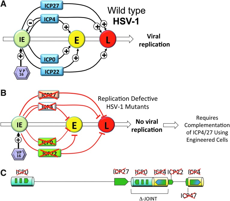
Cascade expression of HSV genes. (A) The viral tegument protein VP16 (purple) activates the IE gene promoters (green) to express the IE gene products (blue) that in turn activate the promoters of the E genes (yellow). The E gene products replicate the viral DNA, releasing expression of the L gene products (red) that package viral genomes de novo and form the virus particle structure. (B)
ICP4 (diploid) and ICP27 are essential IE genes. Expression of E and L genes is blocked in replication-defective HSV deleted for ICP4 and/or ICP27. Vector production requires the use of engineered complementing cells. First-generation replication-defective HSV vectors are deleted for ICP4 (orange). Second-generation vectors are deleted for both ICP4 (orange) and ICP27 (white). Third-generation vectors are functionally deleted for all of the IE genes ICP4, ICP27, ICP0 (diploid) (green), ICP22 (green), and ICP47 (not shown) and are noncytotoxic or interfere with cell division. (C) Genomic structure of the third-generation vector: deletion of the joint sequence and all remaining IE genes blocks the expression of any viral genes requiring complementation of ICP4, ICP27, and ICP0 in engineered U2OS cells.
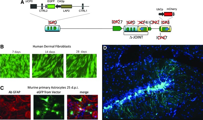
Third-generation replication-defective HSV vector design. (A) Vector deleted for the joint and IE genes with the remaining LAT locus containing an EGFP reporter gene cassette under control of the recombinant C MV enhancer–chicken β-a ctin promoter, splice donor, and intron–human β-g lobin splice acceptor (CAG) promoter. A mCherry reporter gene cassette under control of the ubiquitin C (UbC) promoter is expressed during vector production in complementing cells but silent in noncomplementing cells. (B) EGFP is active long-term in human dermal fibroblasts (28 days); some loss of green cells results from cell division. (C) Mouse astrocytes in culture (25 days); astrocytes are identified using a specific antibody against glial fibrillary acidic protein (GFAP), and in (D) hippocampal neurons after direct intracranial injection using a stereotactic frame (4 days).
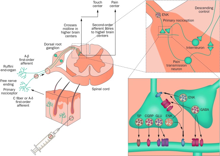
Pain gene therapy using an HSV preproenkephalin-expressing vector. Normal pain signaling pathway control involves signals from peripheral tissues or organs that stimulate the primary first-order afferents that innervate those sites. This leads to the release of various neurotransmitters, including opioid peptides such as ENK, that alter pain signaling to the brain by ascending second-order neurons within the dorsal horn of the spinal cord. The ENK-expressing HSV vector is injected into skin followed by virus uptake into primary afferents; retrograde axonal transport results in HSV vector-mediated production and release of ENK (depicted in orange) from DRG terminals within the dorsal horn. Binding to the specific opioid receptors present on ascending pain transmission neurons and interneurons leads to a block in pain signal transmission. CGRP, calcitonin gene-related peptide; DRG, dorsal root ganglion; ENK, enkephalin; GABA, glutamic acid decarboxylase; GLU, glutamate; SP, substance P. (Figure reproduced with permission from Simonato et al., .)
Similar articles
-
Herpes simplex virus vectors.Mol Cell Biol Hum Dis Ser. 1995;5:33-63. doi: 10.1007/978-94-011-0547-7_3. Mol Cell Biol Hum Dis Ser. 1995. PMID: 9532560 Review. No abstract available.
-
Herpes simplex virus-based vectors.Int J Exp Pathol. 2004 Oct;85(4):177-90. doi: 10.1111/j.0959-9673.2004.00383.x. Int J Exp Pathol. 2004. PMID: 15312123 Free PMC article. Review.
-
Gene delivery and gene therapy with herpes simplex virus-based vectors.Gene. 2001 Feb 7;264(1):1-9. doi: 10.1016/s0378-1119(01)00322-5. Gene. 2001. PMID: 11245972 Review.
-
[Gene therapy for brain tumors using herpes simplex virus vector].Nihon Rinsho. 2005 Sep;63 Suppl 9:510-4. Nihon Rinsho. 2005. PMID: 16201573 Review. Japanese. No abstract available.
-
Replicating herpes simplex virus vectors for cancer gene therapy.Expert Opin Pharmacother. 2000 May;1(4):623-31. doi: 10.1517/14656566.1.4.623. Expert Opin Pharmacother. 2000. PMID: 11249507 Review.
Cited by
-
Current Trends in Viral Gene Therapy for Human Orthopaedic Regenerative Medicine.Tissue Eng Regen Med. 2019 Feb 21;16(4):345-355. doi: 10.1007/s13770-019-00179-x. eCollection 2019 Aug. Tissue Eng Regen Med. 2019. PMID: 31413939 Free PMC article. Review.
-
Efficacy of Herpes Simplex Virus Vector Encoding the Human Preproenkephalin Gene for Treatment of Facial Pain in Mice.J Oral Facial Pain Headache. 2016 Winter;30(1):42-50. doi: 10.11607/ofph.1512. J Oral Facial Pain Headache. 2016. PMID: 26817032 Free PMC article.
-
Pediatric cancer gone viral. Part I: strategies for utilizing oncolytic herpes simplex virus-1 in children.Mol Ther Oncolytics. 2015;2:15015-. doi: 10.1038/mto.2015.15. Epub 2015 Sep 16. Mol Ther Oncolytics. 2015. PMID: 26436135 Free PMC article.
-
Gene Transfer of Brain-derived Neurotrophic Factor (BDNF) Prevents Neurodegeneration Triggered by FXN Deficiency.Mol Ther. 2016 May;24(5):877-89. doi: 10.1038/mt.2016.32. Epub 2016 Feb 5. Mol Ther. 2016. PMID: 26849417 Free PMC article.
-
Virotherapy: cancer gene therapy at last?F1000Res. 2016 Aug 30;5:F1000 Faculty Rev-2105. doi: 10.12688/f1000research.8211.1. eCollection 2016. F1000Res. 2016. PMID: 27635234 Free PMC article. Review.
References
-
- Chattopadhyay M., Goss J., Wolfe D., et al. (2004). Protective effect of herpes simplex virus-mediated neurotrophin gene transfer in cisplatin neuropathy. Brain 127, 929–939 - PubMed
-
- Fraefel C., Jacoby D.R., and Breakefield X.O. (2000). Herpes simplex virus type 1-based amplicon vector systems. Adv. Virus Res. 55, 425–451 - PubMed
Publication types
MeSH terms
LinkOut - more resources
Full Text Sources
Other Literature Sources
