. 2014 Mar 25:1555:89-96.
doi: 10.1016/j.brainres.2014.01.048. Epub 2014 Feb 3.

Fingolimod exerts neuroprotective effects in a mouse model of intracerebral hemorrhage

Affiliations

Fingolimod exerts neuroprotective effects in a mouse model of intracerebral hemorrhage

Lei Lu et al. Brain Res. .

Abstract

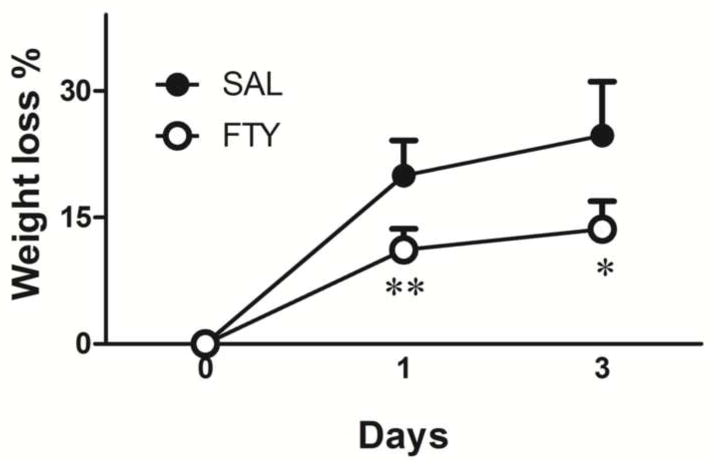
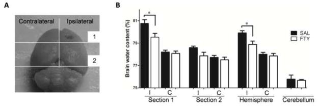
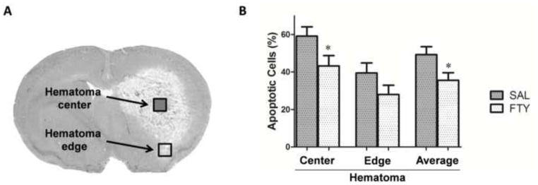
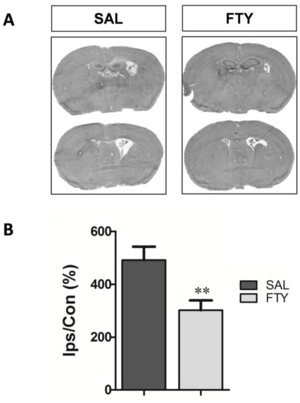
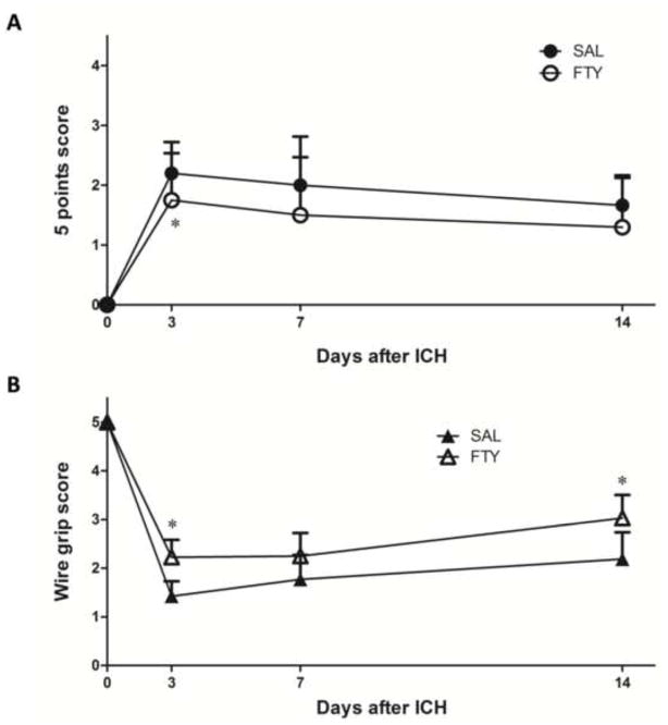
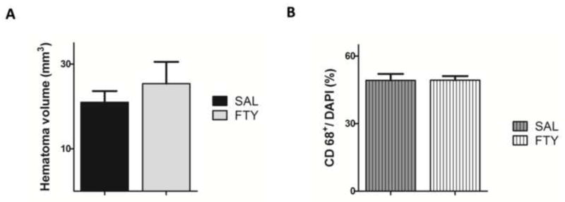
Recent studies have shown that fingolimod (FTY720) is neuroprotective in CNS injury models of cerebral ischemia and spinal cord injury. The purpose of the study was to examine the effect of fingolimod in a mouse model of intracerebral hemorrhage. ICH was produced in adult CD1 mice by injecting collagenase VII-S (0.5 µL, 0.06 U) into the basal ganglia. Fingolimod (or saline) was given 30 min after surgery and once daily for two days. Three days after intracerebral hemorrhage, brain edema, hematoma volume and the number of apoptotic cells were quantified. In another cohort of mice, brain atrophy was evaluated two weeks following intracerebral hemorrhage. Neurobehavioral tests were performed on the 3rd, the 7th and the 14th day. Fingolimod significantly decreased edema, apoptosis and brain atrophy. More importantly, fingolimod enhanced neurobehavioral recovery. Preliminary experiments showed no difference in the number of inflammatory (CD68-positive) cells between the two groups. In conclusion, fingolimod exerts protective effects in a mouse model of intracerebral hemorrhage; the mechanisms underlying these neuroprotective effects deserve further study.

Keywords: FTY720; Intracerebral hemorrhage; Neuroprotection; Sphingosine 1-phosphate; Stroke.


Figures

Fig. 1
Fig. 1. Weight loss after ICH
Compared to pre-surgery, animals lost weight in both treatment groups after collagenase injection. However, fingolimod-treated mice lost less weight than vehicle-treated mice. *p<0.05, **p<0.01, compared to control group, n=10 in each group.
Fig. 2
Fig. 2. Fingolimod reduces brain edema
Brain water content was measured 3 days after ICH in the ipsilateral (I) and contralateral (C) hemispheres. A) Each hemisphere was separated into two sections as shown in Panel A. B) The percentage of water content was expressed as (wet − dry weight) / wet weight. Compared to vehicle-treated mice (SAL), fingolimod (FTY) significantly reduced brain edema. *p<0.05, compared to control group, n=5.
Fig. 3
Fig. 3. Fingolimod decreased apoptotic cells
The apoptotic cells were counted at 3 days after ICH. The ratios of apoptosis were compared between FTY720 and saline group. A) Cells were counted independently in the core of the hemorrhage (open square) and in the peripheral region (filled square). B) Fingolimod significantly reduced the number of apoptotic cells located in the center of the hematoma. Data are presented as mean ± SEM, *p< 0.05, compared to control group, n=10 in each group.
Fig. 4
Fig. 4. Fingolimod decreases brain atrophy
Ventricle sizes were used as indirect measure of brain atrophy 2 weeks after ICH. A) Representative eosin-hematoxylin-stained brain sections in saline (SAL) and fingolimod-treated mice (FTY). B. The ipsilateral (Ips) ventricle size was expressed as a percentage of that of the contralateral (Con). **p< 0.01, compared to control group; n=10 in FTY720 group and n=9 in saline group.
Fig. 5
Fig. 5. Fingolimod improves neurobehavioral functions
Neurobehavioral outcomes were evaluated 3 days, 1 week and 2 weeks after ICH. A) The 5-point scale evaluation was implemented as indicated in the text; higher scores correspond to larger deficits. B) The wire grip performance was quantified as described in Methods; lower scores correspond to larger deficits. FTY=fingolimod, SAL=Saline. *p< 0.05, compared to control group (at 3 days: n=20 in the fingolimod group and n=19 in the saline group; at 1 and 2 weeks, n=10 in the fingolimod group and n=9 in the saline group).
Fig. 6
Fig. 6. Fingolimod does not alter hematoma volume or the number of CD68+ positive cells
A) The volume of hemorrhage was determined using DAB-stained sections. B) The number of CD68 positive cells was compared between the fingolimod and the control groups. Data were presented as mean ± SEM, n=10 in the fingolimod group and n=9 in the saline group.

References

    1. Adeoye O, Broderick JP. Advances in the management of intracerebral hemorrhage. Nat Rev Neurol. 2010;6:593–601. - PubMed
    1. Bermpohl D, You Z, Korsmeyer SJ, Moskowitz MA, Whalen MJ. Traumatic brain injury in mice deficient in Bid: effects on histopathology and functional outcome. J Cereb Blood Flow Metab. 2006;26:625–33. - PubMed
    1. Brinkmann V, Cyster JG, Hla T. FTY720: Sphingosine 1-phosphate receptor-1 in the control of lymphocyte egress and endothelial barrier function. Am J Transplant. 2004;4:1019–1025. - PubMed
    1. Campos F, Qin T, Castillo J, Seo JH, Arai K, Lo EH, Waeber C. Fingolimod reduces hemorrhagic transformation associated with delayed tissue plasminogen activator treatment in a mouse thromboembolic model. Stroke. 2013;44:505–11. - PMC - PubMed
    1. Choi JW, Gardell SE, Herr DR, Rivera R, Lee CW, Noguchi K, Teo ST, Yung YC, Lu M, Kennedy G, Chun J. FTY720 (fingolimod) efficacy in an animal model of multiple sclerosis requires astrocyte sphingosine 1-phosphate receptor 1 (S1P1) modulation. Proc Natl Acad Sci U S A. 2011;108:751–6. - PMC - PubMed

Publication types