. 2014 Feb:450-451:213-21.
doi: 10.1016/j.virol.2013.12.016. Epub 2014 Jan 6.

Orsay virus utilizes ribosomal frameshifting to express a novel protein that is incorporated into virions

Affiliations

Orsay virus utilizes ribosomal frameshifting to express a novel protein that is incorporated into virions

Hongbing Jiang et al. Virology. 2014 Feb.

Abstract

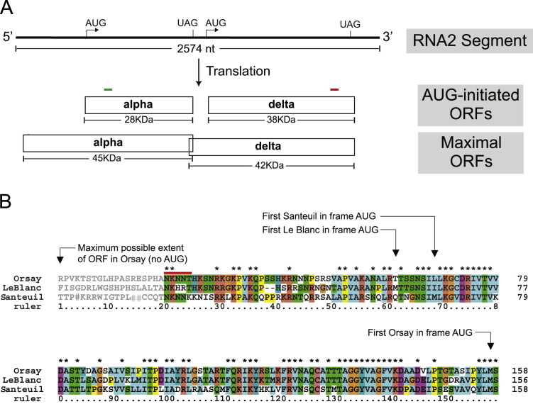
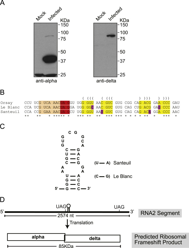
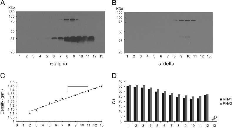
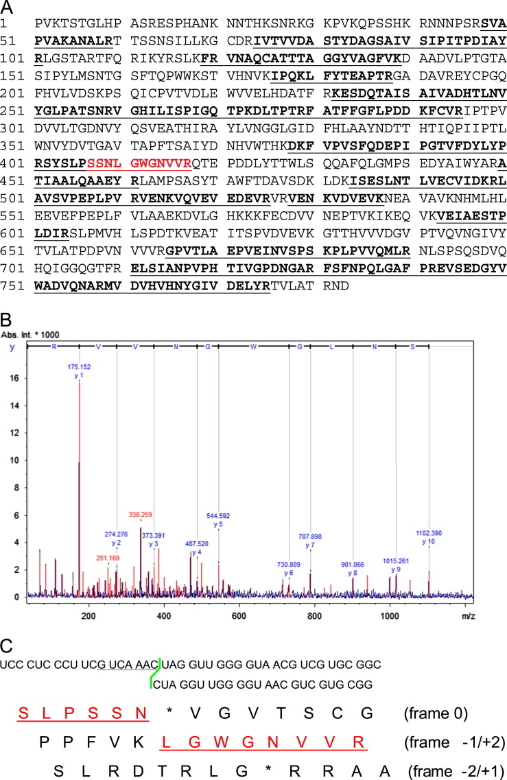
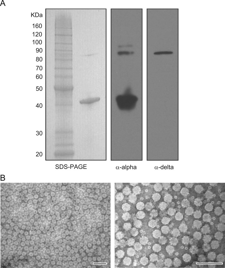
Orsay virus is the first identified virus that is capable of naturally infecting Caenorhabditis elegans. Although it is most closely related to nodaviruses, Orsay virus differs from nodaviruses in its genome organization. In particular, the Orsay virus RNA2 segment encodes a putative novel protein of unknown function, termed delta, which is absent from all known nodaviruses. Here we present evidence that Orsay virus utilizes a ribosomal frameshifting strategy to express a novel fusion protein from the viral capsid (alpha) and delta ORFs. Moreover, the fusion protein was detected in purified virus fractions, demonstrating that it is most likely incorporated into Orsay virions. Furthermore, N-terminal sequencing of both the fusion protein and the capsid protein demonstrated that these proteins must be translated from a non-canonical initiation site. While the function of the alpha-delta fusion remains cryptic, these studies provide novel insights into the fundamental properties of this new clade of viruses.

Keywords: Caenorhabditis elegans; Mass spectrometry; Orsay virus; Ribosomal frameshifting.


Figures

Fig. 1
Fig. 1
Annotation of Orsay virus RNA2 and capsid sequence alignment for three nematode viruses. (A) A schematic model of Orsay virus RNA2 organization and capsid translation initiation. Arrows indicate the first in-frame AUG codon in each ORF. Vertical lines indicate the stop codons. The largest possible ORF that could be generated by a non-AUG start in RNA2 is shown. Green and red bars indicate where the peptide sequences were used to generate alpha and delta peptide derived antibodies, respectively. (B) Alignment of the 5'-proximal regions of RNA2 of all three nematode viruses conceptually translated in the capsid reading frame (‘’ in sequence represents a stop codon). Arrows indicate the first methionine in each sequence. The red bar shows the Orsay virus capsid N-terminal sequence confirmed by sequencing. Sequence in grey shows the conceptual N-terminal translation back to the first in-frame stop codon for Orsay virus. For LeBlanc virus and Santeuil virus, ORF products were also conceptually translated without requiring AUG for initiation. For clarity, the LeBlanc reading frame was truncated to match the length of the maximal Orsay ORF.
Fig. 2
Fig. 2
Western blot of Orsay infected Caenorhabditis elegans suggests possibility of ribosomal frameshifting. (A) Detection of Orsay virus capsid and delta protein in virus infected C. elegans by Western blot using peptide derived antibodies. (B) Alignment of all three nematode virus alpha delta junction regions showed conservation of slippery sequence and stem loop sequence. Spaces separate alpha ORF codons upstream of the shift site (orange) and delta ORF codons downstream of the shift site. The predicted RNA stemloop structure is highlighted with yellow and predicted base-pairings are indicated with parentheses. Paired substitutions that preseve the predicted base-pairings are highlighted with pink. Asterisks indicate conserved positions. (C) The presence of a stem loop structure was modeled by M-fold. Compensatory substitutions are shown for Santeuil and Le Blanc viruses. (D) Predicted model of Orsay virus RNA2 alpha–delta fusion protein translation.
Fig. 3
Fig. 3
CsCl density gradient characterization of Orsay virus. Orsay virus from large scale C. elegans liquid culture was resolved by 20–40% CsCl density gradient. Fractions were numbered 1 to 13 from the top to the bottom of the gradient. (A) Western blot using capsid peptide derived antibody. (B) Western blot using delta peptide derived antibodies. (C) Orsay virus fraction densities. (D) Real time qRT-PCR detection of Orsay virus RNA1 and RNA2. The Y-axis shows the Ct (cycle threshold) value of each fraction. N/D: Ct values were not detected.
Fig. 4
Fig. 4
Mass spectrometry analysis of putative fusion protein. (A) Peptides detected by PMF analysis are highlighted in bold with underline. The peptide sequence spanning the frameshift site is indicated in red. (B) Fragmented peptide ions detected from the peptide spanning the frameshift site in MS/MS analysis. Each charged peptide fragment m/z is shown on the peak. (C) Conceptual translation of the nucleotide sequence spannning the frameshift region in all three reading frames. The red underlined sequence indicates −1 frameshifting product.
Fig. 5
Fig. 5
Iodixanol gradient purification of Orsay virus and electron microscopy study. (A). Coomassie blue staining and Western blot analysis of iodixanol gradient purified Orsay virus. (B). Electron microscopy of iodixanol gradient purified Orsay virions. The structure of purified Orsay virus is shown by negative staining electron microscopy. The scale bar represents 100 nm.

References

    1. Ball L.A., Johnson K.L. Reverse genetics of nodaviruses. Adv. Virus Res. 1999;53:229–244. - PubMed
    1. Brault V., van den Heuvel J.F., Verbeek M., Ziegler-Graff V., Reutenauer A., Herrbach E., Garaud J.C., Guilley H., Richards K., Jonard G. Aphid transmission of beet western yellows luteovirus requires the minor capsid read-through protein P74. EMBO J. 1995;14:650–659. - PMC - PubMed
    1. Brierley I. Ribosomal frameshifting viral RNAs. J. Gen. Virol. 1995;76:1885–1892. - PubMed
    1. Brierley I., Jenner A.J., Inglis S.C. Mutational analysis of the “slippery-sequence” component of a coronavirus ribosomal frameshifting signal. J. Mol. Biol. 1992;227:463–479. - PMC - PubMed
    1. Chao J.A., Lee J.H., Chapados B.R., Debler E.W., Schneemann A., Williamson J.R. Dual modes of RNA-silencing suppression by Flock house virus protein B2. Nat. Struct. Mol. Biol. 2005;12:952–957. - PubMed

Publication types

LinkOut - more resources