. 2014 Apr;111(4):522-532.
doi: 10.1016/j.ymgme.2014.01.008. Epub 2014 Jan 23.

The Pex1-G844D mouse: a model for mild human Zellweger spectrum disorder

Affiliations

The Pex1-G844D mouse: a model for mild human Zellweger spectrum disorder

Shandi Hiebler et al. Mol Genet Metab. 2014 Apr.

Abstract

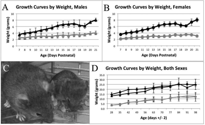
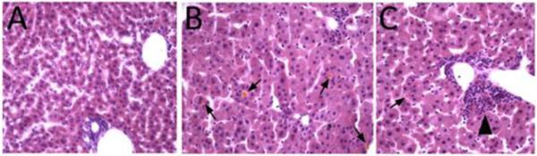
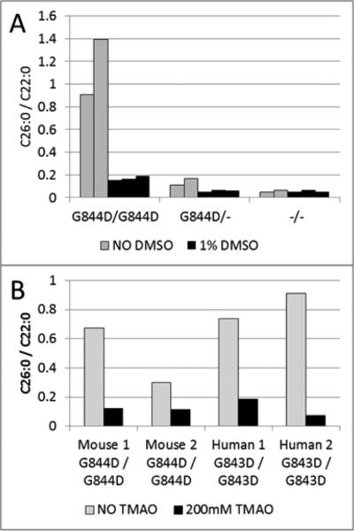
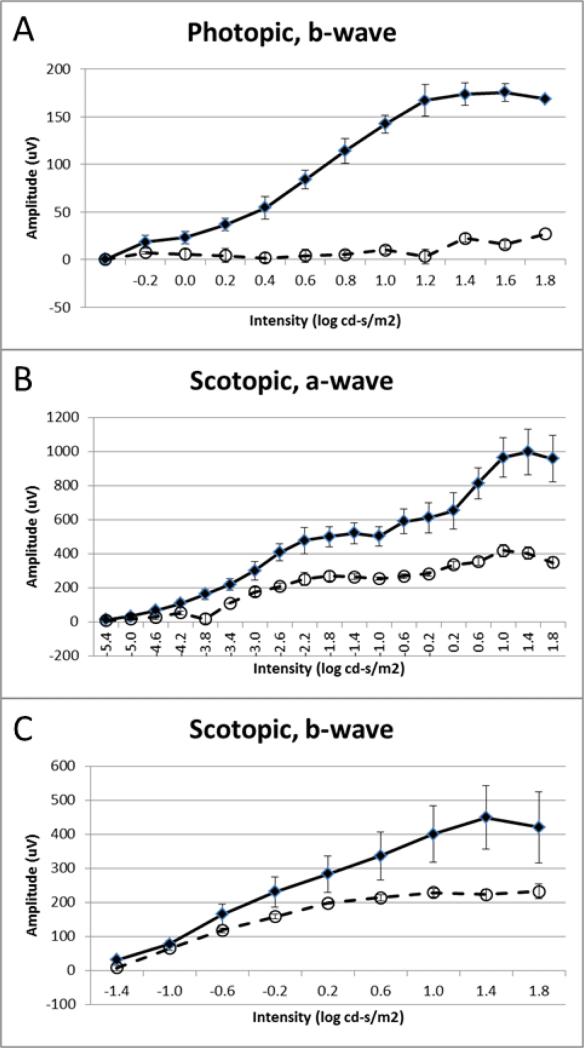
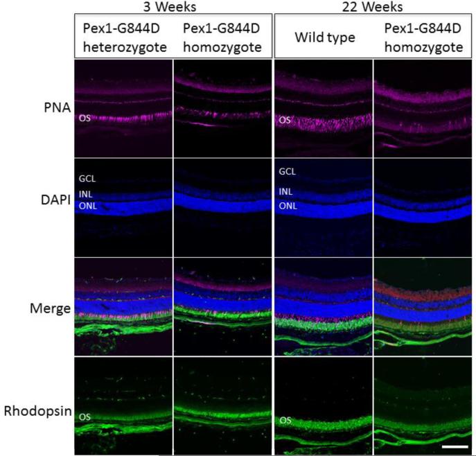
Zellweger spectrum disorder (ZSD) is a disease continuum that results from inherited defects in PEX genes essential for normal peroxisome assembly. These autosomal recessive disorders impact brain development and also cause postnatal liver, adrenal, and kidney dysfunction, as well as loss of vision and hearing. The hypomorphic PEX1-G843D missense allele, observed in approximately 30% of ZSD patients, is associated with milder clinical and biochemical phenotypes, with some homozygous individuals surviving into early adulthood. Nonetheless, affected children with the PEX1-G843D allele have intellectual disability, failure to thrive, and significant sensory deficits. To enhance our ability to test candidate therapies that improve human PEX1-G843D function, we created the novel Pex1-G844D knock-in mouse model that represents the murine equivalent of the common human mutation. We show that Pex1-G844D homozygous mice recapitulate many classic features of mild ZSD cases, including growth retardation and fatty livers with cholestasis. In addition, electrophysiology, histology, and gene expression studies provide evidence that these animals develop a retinopathy similar to that observed in human patients, with evidence of cone photoreceptor cell death. Similar to skin fibroblasts obtained from ZSD patients with a PEX1-G843D allele, we demonstrate that murine cells homozygous for the Pex1-G844D allele respond to chaperone-like compounds, which normalizes peroxisomal β-oxidation. Thus, the Pex1-G844D mouse provides a powerful model system for testing candidate therapies that address the most common genetic cause of ZSD. In addition, this murine model will enhance studies focused on mechanisms of pathogenesis.

Keywords: Bile acids; PEX1; Peroxisome; Photoreceptor degeneration; Retinopathy; Zellweger spectrum disorder.


Figures

Figure 1
Figure 1
Growth from 7 to 98 Days Postnatal. For panels A, B and D, circles denote males, triangles denote females; black denotes control mice, grey denotes Pex1-G844D homozygotes. Panels A and B show the weight in grams of pups from 7 to 21 days postnatal for males (average n = 23 controls and 10 homozygotes) and females (average n = 30 controls and 7 homozygotes), respectively. Panel C shows 21-day-old littermates and highlights the significant growth retardation observed in homozygotes. The mouse on the left is a Pex1-G844D heterozygote and on the right is a Pex1-G844D homozygote and highlights the significant growth retardation observed in homozygotes. Panel D shows the weight in grams of mice from 28 to 98 days postnatal. Males (average n = 13 controls and 5 homozygotes) and females (average n = 14 controls and 4 homozygotes) are shown together.
Figure 2
Figure 2
Liver Histology at 15 Days Postnatal. Hematoxylin-eosin staining. (A) Control mouse. (B, C) Pex1-G844D homozygous mouse. Hepatocyte cytoplasmic volume is increased, associated with microvesicular fat deposition in mutant mice. Yellowish cholestatic deposits (arrows in B, C) are observed in mutant livers, and bile ductular proliferation is seen (arrowhead in C) indicating response to bile duct damage.
Figure 3
Figure 3
Chaperone-molecule Response Testing in Cultured Skin Fibroblasts. Panel A shows results from three mouse fibroblast cell lines representing the three possible genotypes. Each bar in the graph represents the result from a single T25 flask. The homozygous cells treated with 1% DMSO (black bars) for seven days normalized levels of total lipid very long chain fatty acids (VLCFA) based on the C26/C22 ratio as compared to the untreated cells (grey bars). G844D homozygotes are designated G844D/G844D; G844D heterozygotes are designated G844D/-; and wild type is designated −/−. Panel B shows the results from dermal fibroblasts derived from two different Pex1-G844D homozygous mice and two human patients homozygous for PEX1-G843D. The mouse cell lines normalized VLCFA levels after treatment with 200mM TMAO (black bars); this was the same response as PEX1-G843D homozygous human fibroblasts treated simultaneously. Each bar in the graph represents the result from a single T25 flask.
Figure 4
Figure 4
Electrophysiological Functional Analysis of Cone and Rod Visual Systems. ERG measurements were recorded in Pex1-G844D homozygous (open circles, dashed line; n=4) and wild type (black diamonds, solid line; n=4) mice at about two-months of age. A. The amplitudes of photopic b-waves. B. The amplitudes of scotopic a-waves. C. The amplitudes of scotopic b-waves. All data represented as mean ± standard error of the mean. Overall, these data indicate that the cone visual system is significantly impaired in Pex1-G844D homozygotes, whereas the rod photoreceptors are relatively preserved.
Figure 5
Figure 5
Retinal Histology. Immunostaining of photoreceptor outer segments was performed for the Pex1-G884D homozygote and control mouse retinas at three-weeks and 22-weeks. Peanut agglutinin (PNA; magenta) and an antibody against rhodopsin (green) were used to stain cone and rod outer segments, respectively. Nuclei were counter-stained with DAPI (blue). Retinal layers are marked by these abbreviations: GCL, ganglion cells layer; INL, inner nuclear layer; ONL, outer nuclear layer; and, OS, outer segment. Scale bar equals100 mm. Immunostaining with the cone specific PNA in 3-weeks-old mice shows that some cone photoreceptors are preserved in Pex1-G844D homozygotes. In contrast, at 22-weeks of age PNA staining in the Pex1-G844D homozygotes is consistent with a loss of cone photoreceptors. The rhodopsin staining was not as robust in the Pex1-G844D homozygotes as the control indicating that the retinal degeneration may not be limited to the cone photoreceptors.

References

    1. Wanders RJ, Waterham HR. Biochemistry of mammalian peroxisomes revisited. Annual review of biochemistry. 2006;75:295–332. - PubMed
    1. Braverman NE, D'Agostino MD, Maclean GE. Peroxisome biogenesis disorders: Biological, clinical and pathophysiological perspectives. Developmental disabilities research reviews. 2013;17:187–196. - PubMed
    1. Braverman N, Steel G, Obie C, Moser A, Moser H, Gould SJ, Valle D. Human PEX7 encodes the peroxisomal PTS2 receptor and is responsible for rhizomelic chondrodysplasia punctata. Nature genetics. 1997;15:369–376. - PubMed
    1. Ebberink MS, Mooijer PA, Gootjes J, Koster J, Wanders RJ, Waterham HR. Genetic classification and mutational spectrum of more than 600 patients with a Zellweger syndrome spectrum disorder. Human mutation. 2011;32:59–69. - PubMed
    1. Ebberink MS, Koster J, Visser G, Spronsen F, Stolte-Dijkstra I, Smit GP, Fock JM, Kemp S, Wanders RJ, Waterham HR. A novel defect of peroxisome division due to a homozygous non-sense mutation in the PEX11beta gene. Journal of medical genetics. 2012;49:307–313. - PubMed

Publication types

MeSH terms