Review
. 2014 Mar;39(3):101-11.
doi: 10.1016/j.tibs.2013.12.006. Epub 2014 Feb 3.

Many players in BCL-2 family affairs

Affiliations
Review

Many players in BCL-2 family affairs

Tudor Moldoveanu et al. Trends Biochem Sci. 2014 Mar.

Abstract

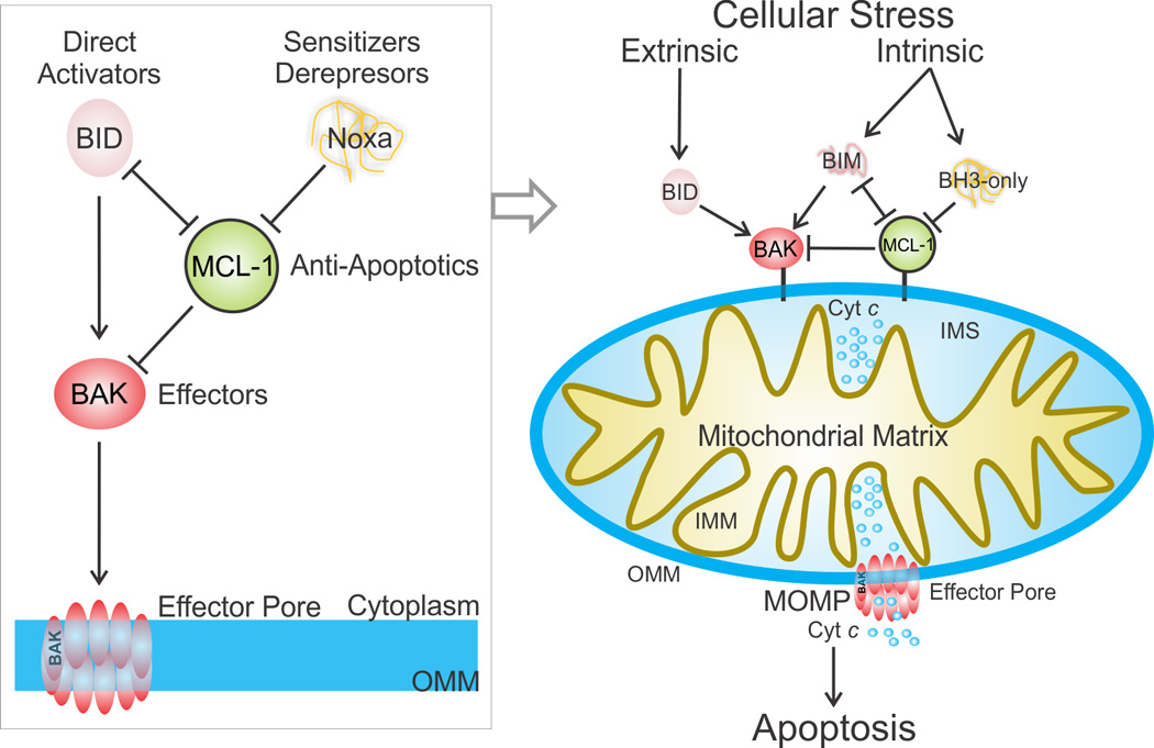
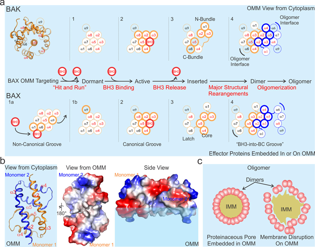
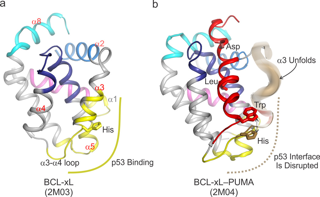
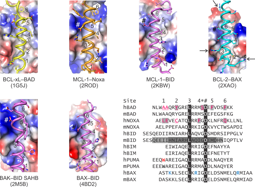
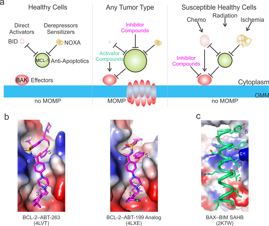
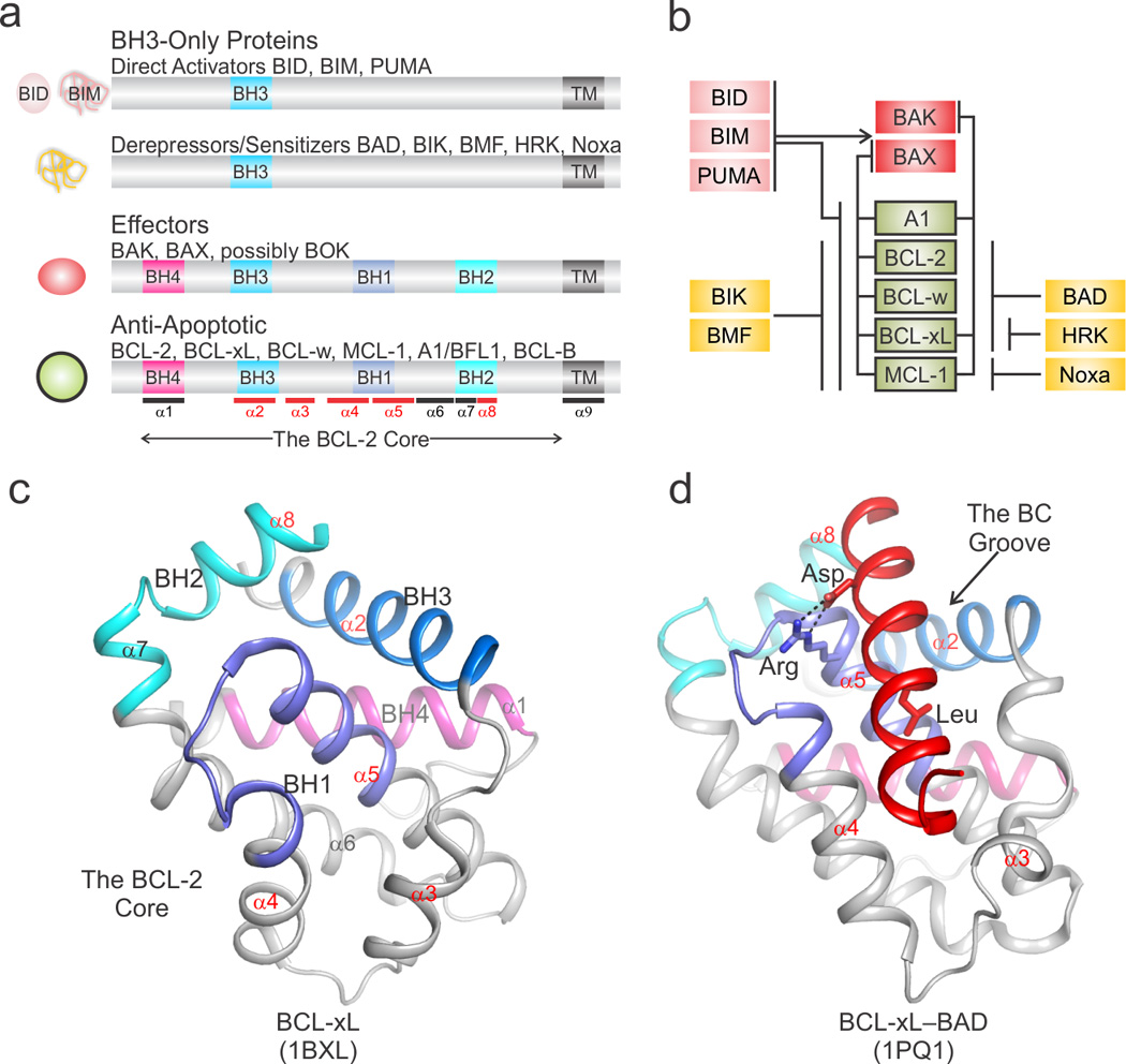
During apoptotic cell death, cellular stress signals converge at the mitochondria to induce mitochondrial outer-membrane permeabilization (MOMP) through B cell lymphoma-2 (BCL-2) family proteins and their effectors. BCL-2 proteins function through protein-protein interactions, the mechanisms and structural aspects of which are only now being uncovered. Recently, the elucidation of the dynamic features underlying their function has highlighted their structural plasticity and the consequent complex thermodynamic landscape governing their protein-protein interactions. These studies show that canonical interactions involve a conserved, hydrophobic groove, whereas non-canonical interactions function allosterically outside the groove. We review the latest structural advances in understanding the interactions and functions of mammalian BCL-2 family members, and discuss new opportunities to modulate these proteins in health and disease.

Keywords: B cell lymphoma-2 (BCL-2) family proteins; mitochondrial apoptosis; mitochondrial outer-membrane permeabilization (MOMP).


Figures

Figure 1
Figure 1. The BCL-2 family protein-mediated mitochondrial pathway of apoptosis
Cell extrinsic and intrinsic cellular stress engages mitochondrial apoptosis through BCL-2 family proteins, which regulate mitochondrial integrity, as shown on the right side of the figure. The extrinsic stress pathway is mounted by ligand binding of death receptors at the plasma membrane resulting in caspase 8-mediated activation of BID (not shown). The intrinsic pathway is poorly characterized in different cell types upstream of BH3-only proteins, and depending on the stress its engagement results in heterogeneous upregulation of BH3-only activities. For example, BIM is upregulated in response to kinase inhibitors and microtubule stabilizing drugs [103]. When the stress activates enough of the pro-apoptotic BH3-only proteins to overcome the anti-apoptotic response, the effectors BAK and BAX are directly activated by BID, BIM, and potentially other BH3-only proteins, and form pores in the outer mitochondrial membrane (OMM) releasing cytochrome c (cyt c), a process known as the mitochondrial outer membrane permeabilization (MOMP). Cyt c triggers the activation of the downstream caspase cascade leading to apoptosis (not shown). The unified model of BCL-2 family protein function in MOMP is depicted at the left. IMM, inner mitochondrial membrane. IMS, inter membrane space. See also Box 1 for a detailed description of BCL-2 family proteins.
Figure 2
Figure 2. Mechanisms of effector-mediated MOMP
a) The effectors undergo a multistep activation mechanism culminating in oligomerization as illustrated in the flow diagram. At least four and five distinct conformations (numbered 1–4 and separated by dotted lines) have been associated with BAK and BAX during MOMP, respectively. The cartoon representation of the structure of the BCL-2 core of BAK (helices α1–α8) at the top left orients the initial model for both BAK and BAX by centering the core on helix α5. BAK is constitutively targeted to the OMM by α9 (conformation 1), and BAX resides primarily in the cytosol (conformation 1a). During apoptosis, direct activator BH3s (red circle) allosterically engage the non-canonical groove of BAX, releasing α9 from the BC groove (the helices that comprise the BC groove are labeled in red font, i.e. α2–α5, α8) on the opposite face and facilitating α9 OMM targeting (conformation 1b). Conformations 1 of BAK and 1b of BAX are considered dormant, as the OMM insertion of helix α9 does not lead to MOMP. Both effectors are directly activated by BH3 engagement at the canonical BC groove (conformation 2). Possible mechanisms for effector conformational changes associated with monomer insertion in OMM include a N- and C-terminal bundle separation for BAK, and a latch and core separation for BAX (conformation 3). Both effectors form seemingly symmetric dimers by a BH3 interaction of one monomer to the canonical BC groove of another (conformation 4). A second unstable interface may be mediated by α6 (indicated by black and blue brackets). Helices actively involved in the step-wise mechanism are shown with a thick outline. Inserted helices are colored light blue like the OMM. OMM is represented by the light blue background. b) A low-resolution crystal structure of the “BH3-into-groove” dimer has suggested that a hydrophobic region (shown in white) may be formed (view from the OMM ) that has the potential to destabilize the outer leaflet of the OMM (side view). Electropositive, electronegative, and neutral (hydrophobic) patches are blue, red, and white, respectively. c) Effector oligomers (pink), made up of conformation 4 dimers in panel a, may form a well-organized proteinaceous pore that perforates the OMM (left), or they may loosely associate and, through the disruption of the OMM outer leaflet, induce destabilization and OMM rupture (right). IMM, inner mitochondrial membrane.
Figure 3
Figure 3. Allosteric regulation of BCL-xL–p53 complex formation by the BH3-only protein PUMA
a) The basic DNA binding domain of p53 binds an acidic lobe of BCL-xL (shown in yellow) comprising the C terminal segment of α1, α3, the loop between α3 and α4, and the C terminus of α5. This non-canonical binding site lies outside the BC groove of BCL-xL (the helices that comprise the BC groove are labeled in red font, i.e. α2–α5, α8) The four BH regions are colored according to the scheme in Figure I (Box1). b) Upon binding of PUMA BH3 (colored red) to the BC groove of BCL-xL, a π-stacking interaction between a tryptophan in PUMA and a histidine in BCL-xL induces unfolding of its α3 (colored brown) and disrupts the interface with p53. Since a tryptophan residue is only found at that particular position in PUMA, and no other BH3 domains, this regulatory mechanism uniquely confers an additional level of signaling complexity to the PUMA–BCL-xL interaction that is absent in other pairs of interacting BCL-2 family proteins.
Figure 4
Figure 4. BC groove specificity for BH3-only ligands
The electrostatic potential on the surface of BC grooves reveals the similarities and differences between folded BCL-2 family proteins, which ultimately dictate the specificity of the binding pairs as summarized in Box 1. Sequence homology highlights features shared by BH3 ligands, including up to seven hydrophobic residues that engage six pockets along the BC groove (labeled 1–6 in the sequence alignment and in the structures) and a conserved Asp always found in a salt bridge (labeled *) preceded by a small residue (labeled #). Peripheral to these positions, which are marked in each structure panel, there are additional interactions, many electrostatic in nature, that contribute to the specificity. For instance, in the BCL-2–BAX complex three positively charged Lys and Arg residues (blue in the alignment and identified by arrows in the structure) were shown to be essential for the affinity of BAX for BCL-2. In addition to the specificity, the BC grooves have evolved to be selective only for certain BH3s; others typically produce structural clashes when modeled based on the known complexes. Up to four residues in BAD BH3 and Noxa BH3, predicted to clash, had to be replaced to directly activate BAK and BAX. Residues highlighted and underlined in human BAD and NOXA were replaced by those found in human BID BH3 for BAK and BAX activation, respectively. Interestingly, specificity of BID BH3 for BAK and BAX was conferred by minimal BH3 regions that required, respectively, a C- and N-terminal extension to a common core (sequence highlighted by dark gray). Black highlights show 100% sequence conservation. Electropositive, electronegative, and neutral (hydrophobic) patches are blue, red and white, respectively.
Figure 5
Figure 5. Targeting apoptosis via the BCL-2 family in biology and disease
a) Targeted apoptotic therapy addresses the upregulation of anti-apoptotic and the downregulation of activator BCL-2 family proteins in cancer and the aberrant overactivation of the pathways during pathological insults and toxicity related stress. We illustrate this be varying the size of the BCL-2 family protein cartoons to reflect the fluctuations in intracellular levels. For example, in healthy cells the BH3-only protein levels are low and their pro-apoptotic activities are kept in check by anti-apoptotic BCL-2 proteins, overall preventing the direct activator–effector axes from being engaged (left panel). In tumors, the BH3-only proteins are down-regulated and the anti-apoptotic BCL-2 proteins are upregulated, together preventing MOMP. Compounds that mimic the activities of BH3-only proteins can be used to activate the effectors and inhibit the anti-apoptotic BCL-2 proteins (middle panel). On the other hand, compounds that inhibit the effectors may be used to block excessive cell death in healthy tissues susceptible to chemotherapy and radiation therapy or in healthy or diseased tissues prone to ischemic cell death. b) The most promising therapeutics have been designed at Abbvie for the anti-apoptotic BCL-2 family proteins, including ABT-263, which targets both BCL-2 and BCL-xL, and the BCL-2-specific compound ABT-199. Both compounds take advantage of scaffolds that engage the BC groove from the third hydrophobic contact (occupied by a conserved Leu in BH3 ligands) to beyond the fifth. These compounds have also been engineered to explore the electrostatic landscape of the BC groove, including a similar charge interaction to the conserved Arg–Asp (labeled *). c) BH3 mimetics for the non-canonical interaction described for the stabilized alpha helix of BCL-2 protein BIM (SAHB) at a groove on BAX distantly resembling the BC groove have also been described, and either SAHBs or their small molecule mimetics may trigger BAX. Importantly, both BAK- and BAX-mediated MOMP require activation by engagement of their BC grooves, which provides additional opportunities for modulating the mitochondrial pathway with activators or inhibitors. Positive, negative, and neutral (hydrophobic) patches are blue, red, and white, respectively.
BOX1 Figure I
BOX1 Figure I. The structure–function cheat sheet of BCL-2 family proteins
a) Schematic representation of BCL-2 family proteins identifying BCL-2 homology (BH) and transmembrane-targeting (TM) regions with respect to the BCL-2 core. At the left, the cartoons illustrate the structured, globular (rounded) or unstructured, intrinsically disordered (noodle-like) nature of the particular class of BCL-2 family proteins. The approximate position of α-helices in the BCL-2 core is marked at the bottom. Helices that delineate the BC groove are highlighted in red. b) The functional interaction network between pro- and anti-apoptotic BCL-2 family members is color coded as in panel a. c) The structure of the BCL-2 core of BCL-xL identifies the BH regions and α-helices colored as in panel a. d) A representative structure highlighting the BH3-into-BC groove interaction for the complex between BCL-xL and the BH3 region of BAD (shown in red). The conserved Leu and Asp side chains of BAD BH3 are engaged in hydrophobic and electrostatic interactions, respectively, with complementary sites in the BCL-2 core. PDB identifiers are shown at the bottom.

References

    1. Hotchkiss RS, et al. Cell death. N. Engl. J. Med. 2009;361:1570–1583. - PMC - PubMed
    1. Green DR. Apoptotic pathways: ten minutes to dead. Cell. 2005;121:671–674. - PubMed
    1. Jiang X, Wang X. Cytochrome C-mediated apoptosis. Annu. Rev. Biochem. 2004;73:87–106. - PubMed
    1. Enari M, et al. A caspase-activated DNase that degrades DNA during apoptosis, and its inhibitor ICAD. Nature. 1998;391:43–50. - PubMed
    1. Suzuki J, et al. Xk-related protein 8 and CED-8 promote phosphatidylserine exposure in apoptotic cells. Science. 2013;341:403–406. - PubMed

Publication types

MeSH terms

Substances