. 2014 Feb 6:4:3894.
doi: 10.1038/srep03894.

Uterine selection of human embryos at implantation

Affiliations

Uterine selection of human embryos at implantation

Jan J Brosens et al. Sci Rep. .

Abstract

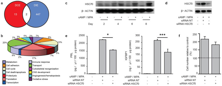
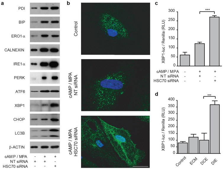
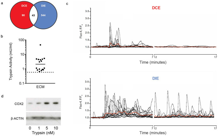
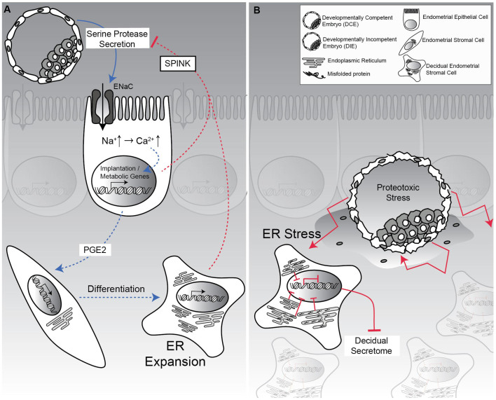
Human embryos frequently harbor large-scale complex chromosomal errors that impede normal development. Affected embryos may fail to implant although many first breach the endometrial epithelium and embed in the decidualizing stroma before being rejected via mechanisms that are poorly understood. Here we show that developmentally impaired human embryos elicit an endoplasmic stress response in human decidual cells. A stress response was also evident upon in vivo exposure of mouse uteri to culture medium conditioned by low-quality human embryos. By contrast, signals emanating from developmentally competent embryos activated a focused gene network enriched in metabolic enzymes and implantation factors. We further show that trypsin, a serine protease released by pre-implantation embryos, elicits Ca(2+) signaling in endometrial epithelial cells. Competent human embryos triggered short-lived oscillatory Ca(2+) fluxes whereas low-quality embryos caused a heightened and prolonged Ca(2+) response. Thus, distinct positive and negative mechanisms contribute to active selection of human embryos at implantation.


Figures

Figure 1
Figure 1. Decidualizing endometrial cells are biosensors of embryo quality.
(a) Venn diagram presenting the number of transcripts regulated in decidualizing HESCs significantly (P < 0.01) regulated in response to signals from developmentally competent embryos (DCE) and developmentally impaired embryos (DIE). (b) Gene Ontology classification of decidual genes regulated in response to soluble factors secreted by DIE. (c) Western blot analysis demonstrating the kinetics of HSC70 induction in primary HESC cultures decidualized with cAMP and MPA in a time-course lasting 8 d. β-ACTIN served as a loading control. A representative result from three different primary cultures is shown. Full length images are presented as Supplementary Information. (d) Primary HESCs were transfected with non-targeting (NT) siRNA or siRNA targeting HSC70, decidualized for 5 d, and then immunoblotted for HSC70. β-ACTIN served as a loading control. Full length images are presented as Supplementary Information. (e) HSC70 knockdown inhibits the secretion of decidual markers, PRL and IGFBP1, in primary HESC cultures differentiated in vitro for 5 d. The data represent mean (±SD) of triplicate experiments. * indicates P < 0.05, and *** P < 0.001. (f) The percentage of viable HESCs, transfected first with non-targeting (NT) siRNA or HSC70 targeting siRNA and then decidualized for 5 d, is presented relative to the number of viable cells in mock-transfected, undifferentiated cells (dotted line). The data represent the mean (±SD) of three biological repeat experiments.
Figure 2
Figure 2. HSC70 knockdown induces ER stress in decidualizing stromal cells.
(a) Total cell lysates from primary HESC cultures, transfected first with non-targeting (NT) siRNA or siRNA targeting HSC70 and then decidualized with cAMP and MPA for 5 d, were immunoprobed for various proteins involved in UPR, ER stress, and autophagy as indicated. β-ACTIN served as a loading control. Full length images are presented as Supplementary Information. (b) HSC70 knockdown in HESCs promotes autophagosome formation. Primary cells cultured on chamber slides were transfected with either non-targeting (NT) or HSC70 targeting siRNA, decidualized for 5 d, stained for LC3B expression (green) and subjected to confocal microscopy. The nuclei were visualized with DAPI (blue). (c) Primary cultures were co-transfected with pcDNA3/XBP1-luc, pRL-sv40 and either siRNA targeting HSC70 or NT siRNA. The cells were left untreated or differentiated for 5 d before measuring luciferase activity. The results show the normalized mean firefly luciferase activity (±SD), expressed in relative light units (RLU), of four biological repeat experiments. *** indicates P < 0.001. (d) Confluent cultures were transfected as described in (c), left untreated (control) or decidualized for 5 d, and then exposed to 30 μl of unconditioned embryo culture medium (ECM) or media conditioned by DCEs or DIEs for 12 h. The results show normalized mean luciferase activity (±SD), expressed in relative light units (RLU), of three biological repeat experiments. *** indicates P < 0.001.
Figure 3
Figure 3. Competent pre-implantation human embryos actively induce a supportive uterine environment.
(a) Venn diagram presenting the number of maternal genes significantly (P < 0.01) altered 24 h after exposure of mouse uterus to unconditioned embryo culture medium or medium conditioned by either developmentally competent or impaired human embryos (DCE and DIE, respectively). (b) Tryptic activity was measured in 16 cultures containing a total of 163 human embryos between day 4 to 6 of development. Activity in unconditioned medium was below dotted line. (c) Application of embryo-conditioned medium induces [Ca2+]i oscillations in Ishikawa cells. Black traces show [Ca2+]i recordings from individual cells in response to application of 1:20 diluted culture medium obtained from DCE (top panel) or DIE (bottom panel). Red traces represent average of all individual traces in each panel. (d) Total protein lysates obtained from Ishikawa cells 24 h after treatment with trypsin (10 nM) for 5 min were subjected to western blot analysis and immunoprobed for COX2. β-ACTIN served as a loading control. Full length images are presented as Supplementary Information.
Figure 4
Figure 4. Positive and negative mechanisms contribute to active selection of human embryos at implantation.
(A) Developmentally competent human embryos secrete evolutionary conserved serine proteases that activate epithelial Na+ channel (ENaC) expressed on luminal epithelial cells, triggering Ca2+ signalling and, ultimately, induction of genes involved in implantation and post-implantation embryo development. In concert, the decidualizing endometrium secretes serine protease inhibitors, such as murine SPINK3 and the human homolog SPINK1, to limit embryo-derived proteolytic activity. Note that acquisition of a secretory phenotype upon decidualization depends on massive expansion of the ER in HESCs. (B) By contrast, excessive protease activity emanating from developmentally compromised embryos that have breached the luminal epithelium down-regulates the expression of molecular chaperones in surrounding decidual cells, leading to accumulation of misfolded proteins and ER stress. This in turn compromises decidual cell functions and triggers tissue breakdown and early maternal rejection.

Similar articles

Cited by

References

    1. Macklon N. S., Geraedts J. P. & Fauser B. C. Conception to ongoing pregnancy: the ‘black box’ of early pregnancy loss. Hum Reprod Update 8, 333–343 (2002). - PubMed
    1. Rai R. & Regan L. Recurrent miscarriage. Lancet 368, 601–611 (2006). - PubMed
    1. Fragouli E. et al. The origin and impact of embryonic aneuploidy. Hum Genet, 10.1007/s00439-013-1309-0 (2013). - PubMed
    1. Mertzanidou A. et al. Microarray analysis reveals abnormal chromosomal complements in over 70% of 14 normally developing human embryos. Hum Reprod 28, 256–264, 10.1093/humrep/des362 (2013). - PubMed
    1. Vanneste E. et al. Chromosome instability is common in human cleavage-stage embryos. Nat Med 15, 577–583 (2009). - PubMed

Publication types

MeSH terms