. 2014 Feb 7;46(2):e75.
doi: 10.1038/emm.2013.147.

Increased expression of the receptor for advanced glycation end products in neurons and astrocytes in a triple transgenic mouse model of Alzheimer's disease

Affiliations

Increased expression of the receptor for advanced glycation end products in neurons and astrocytes in a triple transgenic mouse model of Alzheimer's disease

Bo-Ryoung Choi et al. Exp Mol Med. .

Abstract

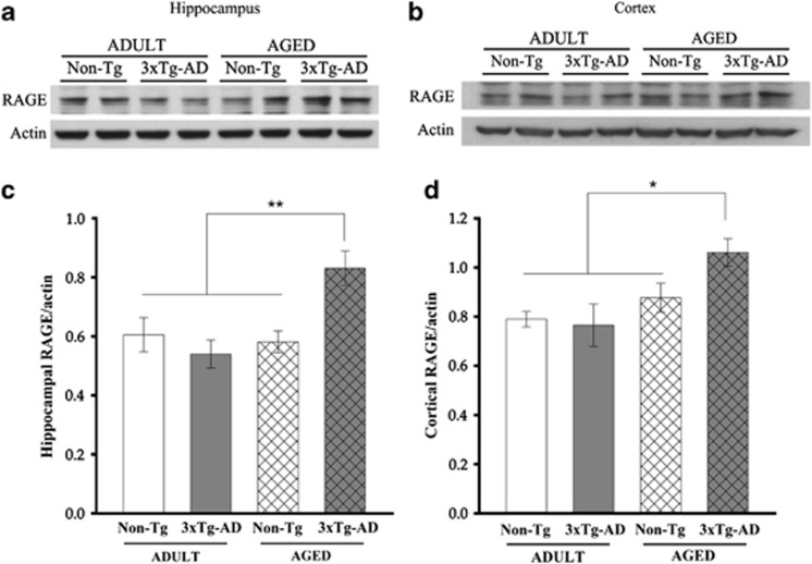
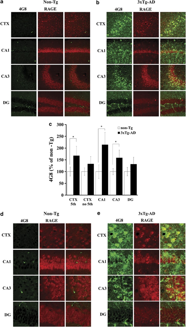
The receptor for advanced glycation end products (RAGE) has been reported to have a pivotal role in the pathogenesis of Alzheimer's disease (AD). This study investigated RAGE levels in the hippocampus and cortex of a triple transgenic mouse model of AD (3xTg-AD) using western blotting and immunohistochemical double-labeling to assess cellular localization. Analysis of western blots showed that there were no differences in the hippocampal and cortical RAGE levels in 10-month-old adult 3xTg-AD mice, but significant increases in RAGE expression were found in the 22- to 24-month-old aged 3xTg-AD mice compared with those of age-matched controls. RAGE-positive immunoreactivity was observed primarily in neurons of aged 3xTg-AD mice with very little labeling in non-neuronal cells, with the notable exception of RAGE presence in astrocytes in the hippocampal area CA1. In addition, RAGE signals were co-localized with the intracellular amyloid precursor protein (APP)/amyloid beta (Aβ) but not with the extracellular APP/Aβ. In aged 3xTg-AD mice, expression of human tau was observed in the hippocampal area CA1 and co-localized with RAGE signals. The increased presence of RAGE in the 3xTg-AD animal model showing critical aspects of AD neuropathology indicates that RAGE may contribute to cellular dysfunction in the AD brain.


Figures

Figure 1
Figure 1
Levels of hippocampal and cortical RAGE in adult and aged 3xTg-AD and non-Tg control mice. Representative immunoblots of RAGE (45 kDa) expression in the hippocampus (a) and cortex (b) in adult and aged 3xTg-AD and non-Tg control mice. Levels of RAGE were significantly higher in aged 3xTg-AD mice than in aged non-Tg control, adult 3xTg-AD and adult non-Tg control mice (c, d). Data are expressed as the ratios of RAGE/actin (means±s.e.m.; n=6 per each group). *P<0.05, **P<0.01.
Figure 2
Figure 2
Distribution and cellular localization of RAGE in aged 3xTg-AD. Immunofluorescent RAGE images in the hippocampus and cortex (CTX) in aged non-Tg control and 3xTg-AD mouse (a). Confocal images of double-labeling with RAGE and NeuN or Iba-1 in the hippocampus and cortex in aged non-Tg control (b) and 3xTg-AD (c) mouse. The immunoreactivity of RAGE and Iba-1-positive microglia were increased in 3xTg-AD mice compared with non-Tg controls (d, e). No differences in NeuN immunoreactivity were observed between these two groups (f). The majority of RAGE labeling overlapped with NeuN-positive neurons, although some labeling was found in non-neuronal cells (c). Data are the means±s.e.m. (adult: n=6; aged: n=10). *P<0.05, **P<0.01. Scale bar=200 μm.
Figure 3
Figure 3
Co-localization of RAGE with GFAP-positive astrocytes in the hippocampal area CA1 of aged 3xTg-AD mice. Confocal images of triple-labeling with RAGE, GFAP and Iba-1 in the hippocampus and cortex (CTX) in aged non-Tg control (a) and 3xTg-AD (b) mouse. Immunoreactivity of GFAP-positive astrocytes in aged 3xTg-AD mice was not significantly different from that in aged non-Tg control mice, even though immunoreactivity in the CA1 was higher (c). However, the signals of RAGE (blue) co-localized with GFAP (green)-positive astrocytes (Iba-1 (red), d) (see reverse triangles). The number of RAGE-expressing astrocytes in the hippocampal area CA1 in aged3xTg-AD was significantly higher than in non-Tg controls (e,*P<0.05). Data are the means±s.e.m. (adult: n=6; aged: n=10). Scale bar=200 μm.
Figure 4
Figure 4
Distribution of 4G8-positive APP/Aβ and its co-localization with RAGE in aged 3xTg-AD mice. Confocal images of double-labeling with RAGE and 4G8 in the hippocampus and cortex (CTX) in aged non-Tg control (a, d) and 3xTg-AD (b, e) mouse. Immunoreactive labeling for 4G8, a monoclonal antibody detecting APP/Aβ, was increased in aged 3xTg-AD mice compared with aged non-Tg controls (c). Notably, in the cortex, the immunoreactivity of 4G8 was significantly increased only in layer 5, whereas there were no differences in other layers (c). In the hippocampus, 4G8 immunoreactivity was significantly increased in area CA1 and CA3 but not in the DG (c). Labeling for intracellular 4G8 in neurons was co-localized with RAGE expression, whereas that for extracellular 4G8 was not (e). Data are means±s.e.m. (adult: n=6; aged: n=10). *P<0.05. Scale bar=100 μm.
Figure 5
Figure 5
Distribution of HT7-positive human tau protein and its co-localization with RAGE in aged 3xTg-AD mice. Confocal images of double-labeling with RAGE and HT7 in the hippocampus and cortex (CTX) in aged non-Tg control (a, d) and 3xTg-AD (b, e) mice. Immunoreactivity of HT7, a marker of human tau protein, was higher in the hippocampal area CA1 of aged 3xTg-AD mice compared with aged non-Tg control (c). HT7 labeling overlapped with RAGE. In aged 3xTg-AD mice, HT7 was expressed in some cortical neurons, CA1 neurons, and some axons of the mossy fiber, whereas the DG and hippocampal area CA3 did not show immunoreactivity for HT7 when compared with aged non-Tg controls (e). Data are means±s.e.m. (adult: n=6; aged: n=10). **P<0.01. Scale bar=100 μm.

References

    1. Mattson MP. Pathways towards and away from Alzheimer's disease. Nature. 2004;430:631–639. - PMC - PubMed
    1. Selkoe DJ. Alzheimer's disease: genes, proteins, and therapy. Physioll Rev. 2001;81:741–766. - PubMed
    1. Billings LM, Oddo S, Green KN, McGaugh JL, LaFerla FM. Intraneuronal Abeta causes the onset of early Alzheimer's disease-related cognitive deficits in transgenic mice. Neuron. 2005;45:675–688. - PubMed
    1. LaFerla FM, Green KN, Oddo S. Intracellular amyloid-beta in Alzheimer's disease. Nat Rev Neurosci. 2007;8:499–509. - PubMed
    1. Oddo S, Caccamo A, Kitazawa M, Tseng BP, LaFerla FM. Amyloid deposition precedes tangle formation in a triple transgenic model of Alzheimer's disease. Neurobiol Aging. 2003;24:1063–1070. - PubMed

Publication types

MeSH terms

Substances

LinkOut - more resources