. 2014 Apr 29;129(17):1770-80.
doi: 10.1161/CIRCULATIONAHA.113.006797. Epub 2014 Feb 6.

Aberrant chloride intracellular channel 4 expression contributes to endothelial dysfunction in pulmonary arterial hypertension

Affiliations

Aberrant chloride intracellular channel 4 expression contributes to endothelial dysfunction in pulmonary arterial hypertension

Beata Wojciak-Stothard et al. Circulation. .

Abstract

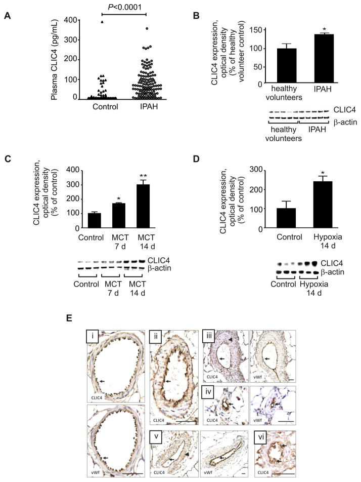
Background: Chloride intracellular channel 4 (CLIC4) is highly expressed in the endothelium of remodeled pulmonary vessels and plexiform lesions of patients with pulmonary arterial hypertension. CLIC4 regulates vasculogenesis through endothelial tube formation. Aberrant CLIC4 expression may contribute to the vascular pathology of pulmonary arterial hypertension.

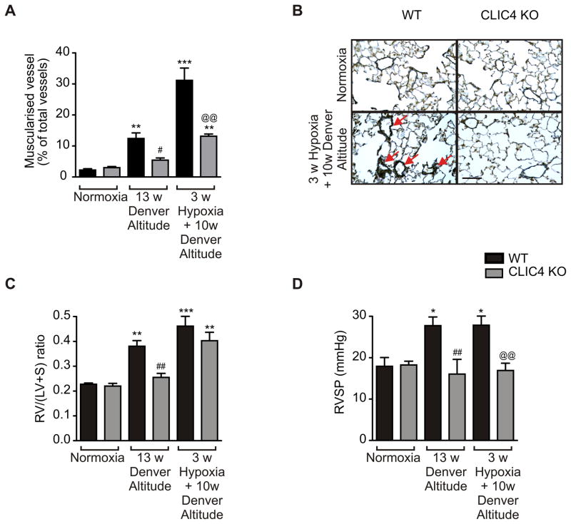
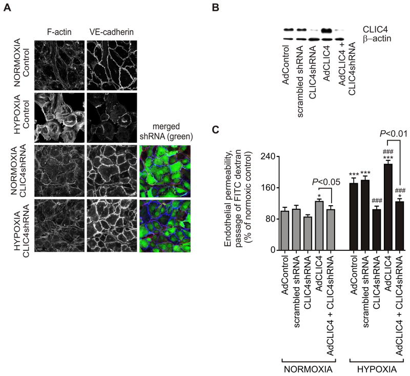
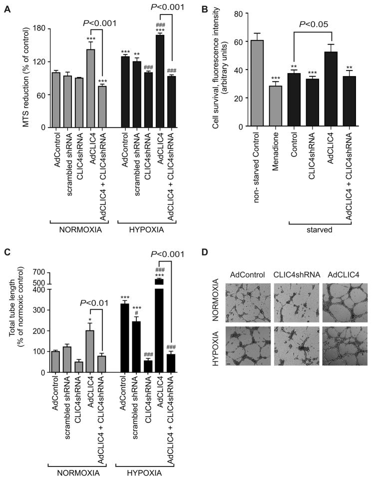
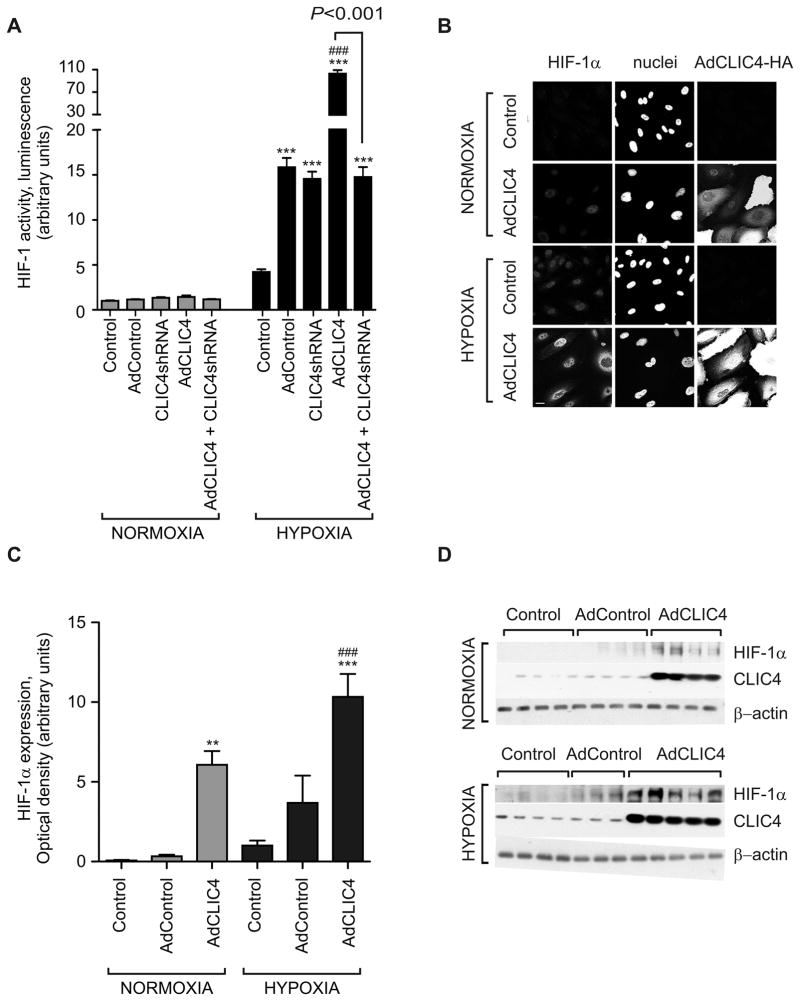
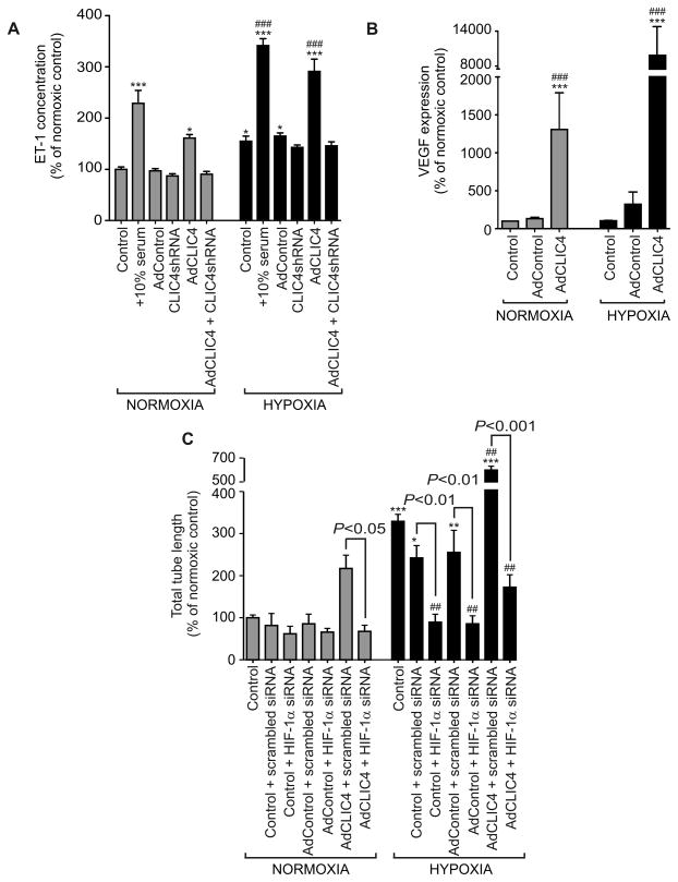
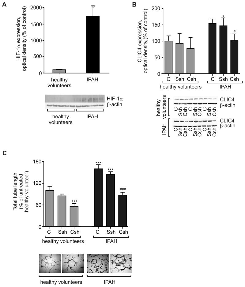
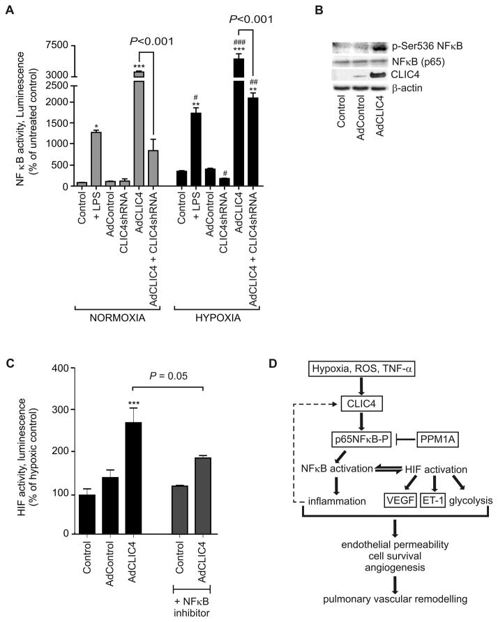
Methods and results: CLIC4 protein expression was increased in plasma and blood-derived endothelial cells from patients with idiopathic pulmonary arterial hypertension and in the pulmonary vascular endothelium of 3 rat models of pulmonary hypertension. CLIC4 gene deletion markedly attenuated the development of chronic hypoxia-induced pulmonary hypertension in mice. Adenoviral overexpression of CLIC4 in cultured human pulmonary artery endothelial cells compromised pulmonary endothelial barrier function and enhanced their survival and angiogenic capacity, whereas CLIC4 shRNA had an inhibitory effect. Similarly, inhibition of CLIC4 expression in blood-derived endothelial cells from patients with idiopathic pulmonary arterial hypertension attenuated the abnormal angiogenic behavior that characterizes these cells. The mechanism of CLIC4 effects involves p65-mediated activation of nuclear factor-κB, followed by stabilization of hypoxia-inducible factor-1α and increased downstream production of vascular endothelial growth factor and endothelin-1.

Conclusion: Increased CLIC4 expression is an early manifestation and mediator of endothelial dysfunction in pulmonary hypertension.

Keywords: angiogenesis inducing agents; endothelium; hypertension, pulmonary; hypoxia-inducible factor 1; nuclear factor-kappaB.


Conflict of interest statement

Conflict of Interest Disclosures: None.

Figures

Figure 1
Figure 1
CLIC4 protein expression in patient plasma, human ECFCs and rat lung. (A) CLIC4 protein levels in plasma samples from IPAH patients (n=123) and healthy volunteers (n=64). As these data show a skewed distribution they were analysed using the Mann-Whitney U-test. (B) Levels determined in blood-derived endothelial cells from IPAH patients (n=4–5). (C) CLIC4 protein expression in the lungs of rats treated with monocrotaline (MCT) or (D) exposed to hypoxia (n= 3–8). Data are presented as mean±SEM. *P<0.05; **P<0.01, compared with controls. Lung sections from (E i) control, (E ii) hypoxia-, (E iii–iv) MCT and (E v–vi) Sugen 5416/hypoxia-induced pulmonary hypertensive rats, demonstrating co-localisation of CLIC4 and von Willebrand factor (vWf) in the pulmonary endothelium (arrows) of normal and remodelled pulmonary arteries. Arrowheads (E iii, v) indicate CLIC4 immunoreactivity in smooth muscle cells. Bar=50 μm.
Figure 2
Figure 2
CLIC4 gene knockout attenuates development of chronic hypoxia-induced pulmonary hypertension. (A) Percentage of muscularized vessels diameter ≤50 μm in lung sections of normoxic and hypoxic wildtype (WT; black bars) and CLIC4 knockout (CLIC4 KO; grey bars) mice. (B) Representative sections showing α-smooth muscle actin staining in normoxic or hypoxic WT or CLIC4 KO mice. Red arrows point to the fully muscularized peripheral artery with double elastic lamina staining. Bar=50 μm. (C) Right ventricular hypertrophy (RV/(LV+S)) and (D) right ventricular systolic pressure (RVSP) were measured in the WT control and CLIC4 KO mice following exposure to normoxia, 13 weeks at Denver altitude or 10 weeks (w) of Denver altitude followed by 3 weeks of hypoxia. *P<0.05, **P<0.01, ***P<0.01, compared to normoxic WT; #P<0.05, ##P<0.01 compared to WT at Denver altitude and @@P<0.01, WT versus CLIC4 KO group at 3w Hypoxia+10w Denver Altitude; (C). In (A–C) n=4–8.
Figure 3
Figure 3
Hypoxia-induced changes in human pulmonary endothelial phenotype and permeability. (A) Images showing the organization of F-actin and VE-cadherin in confluent HPAECs under normoxic or hypoxic (24 hr) conditions. The cells were left untreated or overexpressed CLIC4 shRNA, as indicated. Merged images show F-actin (red), VE-cadherin (blue) and GFP co-expressed with CLIC4 shRNA (green). Bar=10 μm. (B) The adenoviral manipulation of CLIC4 expression was confirmed by western blot analysis of cell extracts. (C) Pulmonary endothelial cell permeability in normoxic and hypoxic cells expressing scrambled shRNA, CLIC4 shRNA or CLIC4, as indicated; In (C) data are expressed as % of control and each bar represents mean±SEM (n=4). *P<0.05; ***P<0.001, compared with normoxic AdControl; ###P<0.001, compared with hypoxic AdControl. Comparisons between AdCLIC4 and CLIC4 shRNA + AdCLIC4 are also indicated.
Figure 4
Figure 4
Effects of CLIC4 expression on HPAEC metabolic activity, cell survival and angiogenesis. (A) Cell metabolic activity under normoxic and hypoxic conditions and following overexpression of CLIC4, scrambled shRNA (shRNA control) or CLIC4 shRNA, as indicated; MTS reduction assay. (B) HPAEC viability in cells cultured in full media, treated with apoptosis-inducer, menadione (6 hr, 10 μM) or serum-starved for 48 hr. The cells were overexpressing CLIC4 or CLIC4 and CLIC4 shRNA, as indicated. (C) Changes in total endothelial tube length (expressed as % of normoxic control) in cells treated, as indicated. (D) Representative images showing the effects AdCLIC4 and AdCLIC4 shRNA on tube formation in normoxic and hypoxic HPAECs. Bar=500 μm. In (A–C) each bar represents mean±SEM; (n=4–5). *P<0.05, **P<0.01, ***P<0.001, compared with normoxic control (A–C) or non-starved control (B); ###P<0.001, compared with hypoxic AdControl (A, C). Comparisons between AdCLIC4 and AdCLIC4 + CLIC4 shRNA groups are also indicated.
Figure 5
Figure 5
CLIC4 regulates endothelial HIF activity and stabilization. Overexpression of CLIC4 (24 hr) or exposure to hypoxia (A) stabilizes HIF in U2OS-HRE-luc cells, luciferase reporter assay (n=12); (B) increases nuclear translocation of HIF-1α and (C) increases HIF-1α levels in HPAECs (n=4–5). Each bar represents mean±SEM ***P<0.001, **P<0.01, compared with normoxic AdControl; ###P<0.001, compared with hypoxic AdControl. Comparisons between AdCLIC4 and CLIC4 shRNA + AdCLIC4, are also indicated
Figure 6
Figure 6
The effect of CLIC4 on HIF-dependent protein expression and tube formation in HPAECs. Production of (A) ET-1 and (B) VEGF in normoxic and hypoxic HPAECs overexpressing CLIC4 or CLIC4 shRNA (n=3–5). (C) HPAEC tube formation following siRNA-mediated HIF-1α knockdown in cells treated, as indicated (n=5). Each bar represents mean±SEM. *P<0.05; **P<0.01, ***P<0.001 compared with normoxic AdControl; ##P<0.01, ###P<0.001 compared with hypoxic AdControl. Comparisons between AdCLIC4 and CLIC4 shRNA or between scrambled siRNA and HIF-1α siRNA groups are also indicated.
Figure 7
Figure 7
HIF-1α levels and tube formation in blood-derived endothelial cells. (A) Blood-derived endothelial cells from IPAH patients showed greater protein expression of HIF-1α compared to healthy controls. (B) CLIC4 shRNA (CSh; 48h) reduces CLIC4 levels in IPAH cells to control levels, while scrambled shRNA (Ssh) has no effect. (C) CLIC4 shRNA inhibits tube formation in IPAH endothelial cells. Each bar represents mean±SEM (n=3–4). Representative images of tube formation in healthy and IPAH cells are shown below the graph; Bar=100 μm. In (A–D) *P<0.05, **P<0.01, ***P<0.001, compared with untreated cells derived from healthy volunteers. #P<0.05, ###P<0.01 comparison with untreated IPAH cells.
Figure 8
Figure 8
CLIC4 regulates NFκB activity in HPAECs. (A) NFκB activity in HPAECs treated with lipopolysacharide (LPS), control adenovirus (AdControl), AdCLIC4 or AdCLIC4 shRNA in normoxic (grey bars) or hypoxic (black bars) conditions, as indicated; luciferase reporter assay. (B) CLIC4 increases phosphorylation of p65 on Serine 536, western blotting. (C) NFκB inhibitor, BAY 117085 prevents HIF activation in CLIC4- overexpressing HPAECs (30 hr overexpression) in hypoxia. n=4; *P<0.05; **P<0.01, ***P<0.001, compared with untreated controls; #P<0.05, ##P <0.01, ###P <0.001, comparison with hypoxic controls. (D) The proposed CLIC4 signalling pathway.

References

    1. Littler DR, Harrop SJ, Goodchild SC, Phang JM, Mynott AV, Jiang L, Valenzuela SM, Mazzanti M, Brown LJ, Breit SN, Curmi PM. The enigma of the CLIC proteins: Ion channels, redox proteins, enzymes, scaffolding proteins? FEBS Lett. 2010;584:2093–2101. - PubMed
    1. Bohman S, Matsumoto T, Suh K, Dimberg A, Jakobsson L, Yuspa S, Claesson-Welsh L. Proteomic analysis of vascular endothelial growth factor-induced endothelial cell differentiation reveals a role for chloride intracellular channel 4 (CLIC4) in tubular morphogenesis. J Biol Chem. 2005;280:42397–42404. - PubMed
    1. Tung JJ, Hobert O, Berryman M, Kitajewski J. Chloride intracellular channel 4 is involved in endothelial proliferation and morphogenesis in vitro. Angiogenesis. 2009;12:209–220. - PMC - PubMed
    1. Chalothorn D, Zhang H, Smith JE, Edwards JC, Faber JE. Chloride intracellular channel-4 is a determinant of native collateral formation in skeletal muscle and brain. Circ Res. 2009;105:89–98. - PMC - PubMed
    1. Abdul-Salam VB, Wharton J, Cupitt J, Berryman M, Edwards RJ, Wilkins MR. Proteomic analysis of lung tissues from patients with pulmonary arterial hypertension. Circulation. 2010;122:2058–2067. - PubMed

MeSH terms