. 2014 Feb 4;9(2):e86939.
doi: 10.1371/journal.pone.0086939. eCollection 2014.

Increased anatomical specificity of neuromodulation via modulated focused ultrasound

Affiliations

Increased anatomical specificity of neuromodulation via modulated focused ultrasound

Edin Mehić et al. PLoS One. .

Abstract

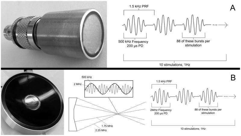
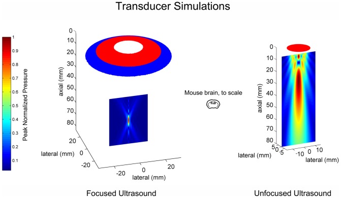
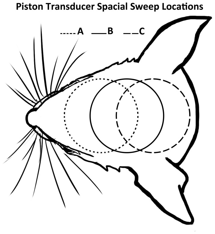
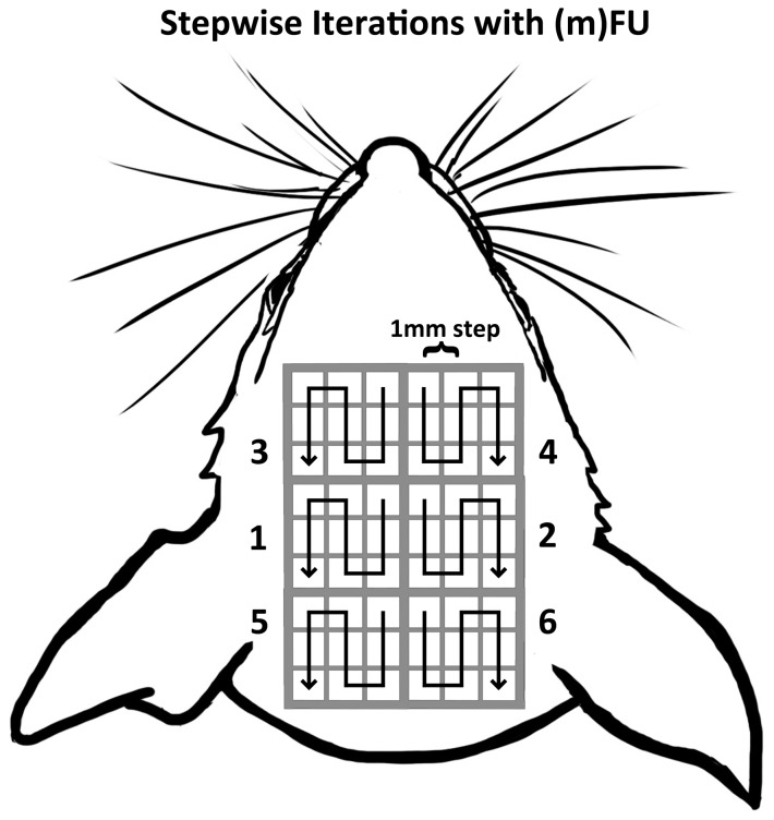
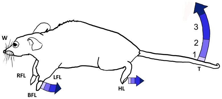
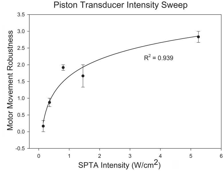
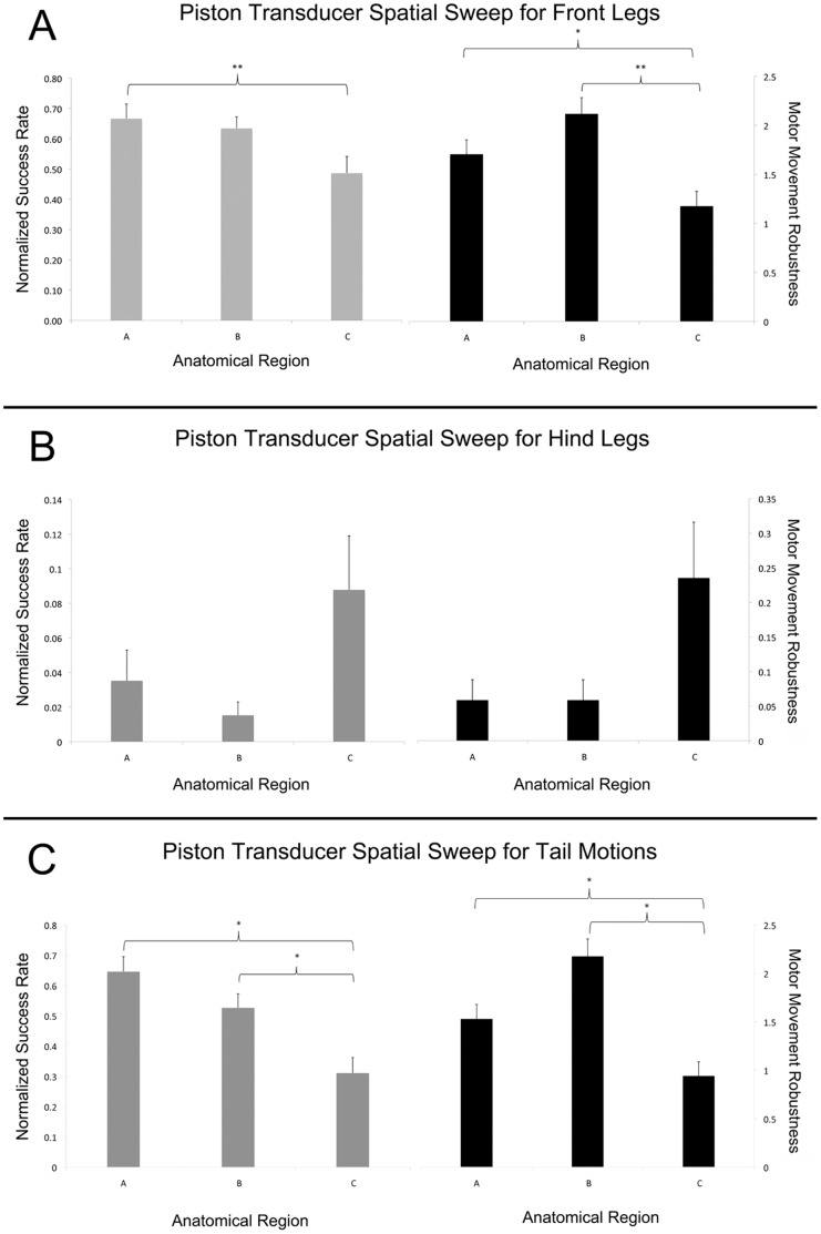
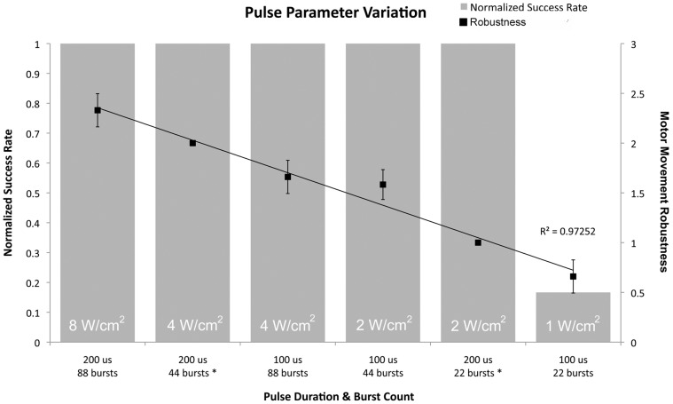
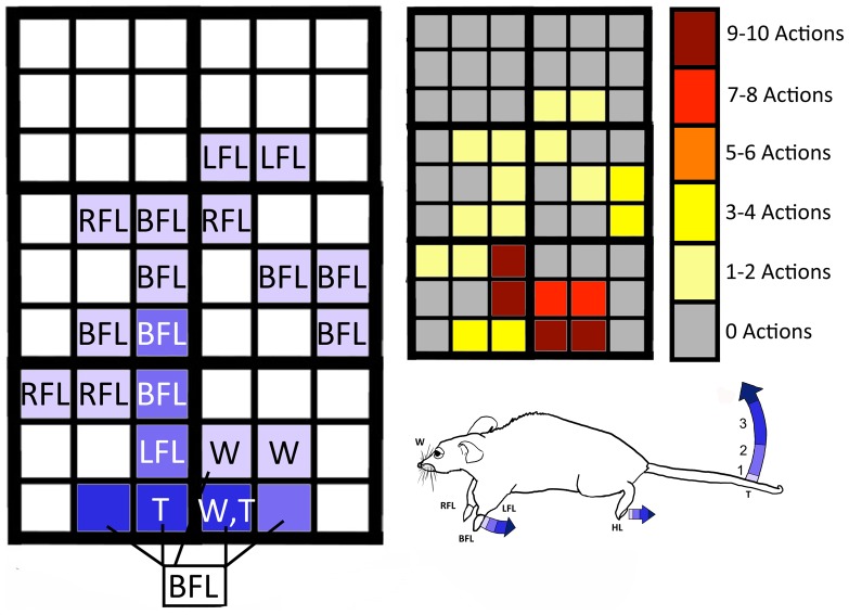
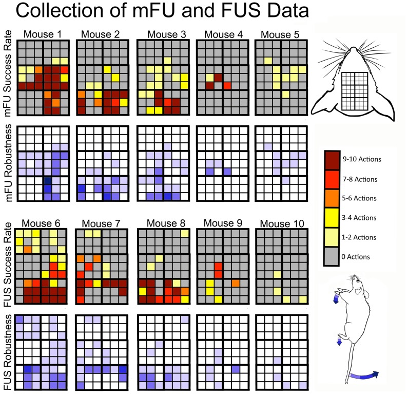
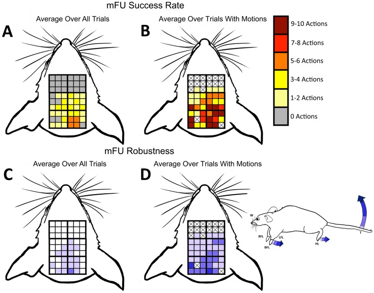
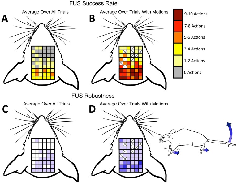
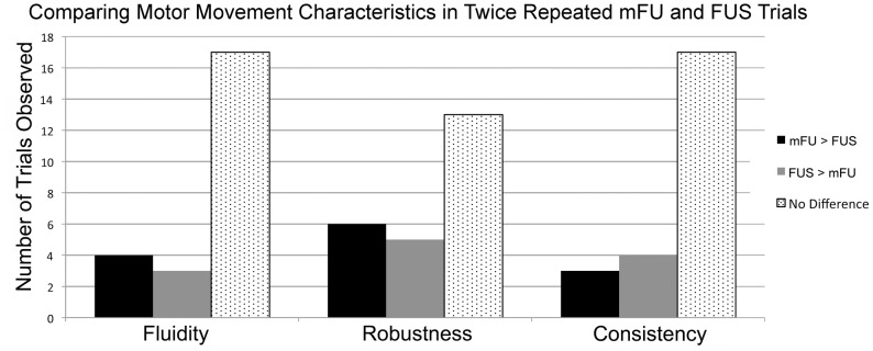
Transcranial ultrasound can alter brain function transiently and nondestructively, offering a new tool to study brain function now and inform future therapies. Previous research on neuromodulation implemented pulsed low-frequency (250-700 kHz) ultrasound with spatial peak temporal average intensities (ISPTA) of 0.1-10 W/cm(2). That work used transducers that either insonified relatively large volumes of mouse brain (several mL) with relatively low-frequency ultrasound and produced bilateral motor responses, or relatively small volumes of brain (on the order of 0.06 mL) with relatively high-frequency ultrasound that produced unilateral motor responses. This study seeks to increase anatomical specificity to neuromodulation with modulated focused ultrasound (mFU). Here, 'modulated' means modifying a focused 2-MHz carrier signal dynamically with a 500-kHz signal as in vibro-acoustography, thereby creating a low-frequency but small volume (approximately 0.015 mL) source of neuromodulation. Application of transcranial mFU to lightly anesthetized mice produced various motor movements with high spatial selectivity (on the order of 1 mm) that scaled with the temporal average ultrasound intensity. Alone, mFU and focused ultrasound (FUS) each induced motor activity, including unilateral motions, though anatomical location and type of motion varied. Future work should include larger animal models to determine the relative efficacy of mFU versus FUS. Other studies should determine the biophysical processes through which they act. Also of interest is exploration of the potential research and clinical applications for targeted, transcranial neuromodulation created by modulated focused ultrasound, especially mFU's ability to produce compact sources of ultrasound at the very low frequencies (10-100s of Hertz) that are commensurate with the natural frequencies of the brain.


Conflict of interest statement

Competing Interests: The authors have declared that no competing interests exist.

Figures

Figure 1
Figure 1. Transducers and their associated ultrasound emissions.
(A) Ultran planar ultrasound transducer with corresponding waveform representation. (B) Sonic Concepts focused ultrasound transducer (black annulus with filled hole) with corresponding waveform representation.
Figure 2
Figure 2. Ultrasound pressure fields for our devices.
Simulations of focused and planar ultrasound beam plots, with mouse brain for comparison.
Figure 3
Figure 3. Superficial projection of intra-cranial stimulation regions crated by planar ultrasound.
Three regions of quantitatively different stimulation responses created by a sweep of our planar ultrasound device.
Figure 4
Figure 4. Superficial projection of intra-cranial stimulation regions created by mFU and FUS.
The 54 squares represent the superficial projection of individual, intra-cranial stimulation regions, with centers separated in 1 mm increments. Trials began in region 1 and concluded in region 6, following the arrow within each region.
Figure 5
Figure 5. Ordinal robustness scale for motor movements.
A value of one represents minimally observable motion while a value of three represents the largest motions regularly observed.
Figure 6
Figure 6. Intensity of mFU stimulation versus robustness of associated observed motor movement.
Curve represents a logarithmic fit. (N = 3).
Figure 7
Figure 7. Success rate and robustness of movements induced by ultrasound from a planar source.
We report these values for (A) for front legs (B) for hind legs and (C) for tail. The success rate was normalized to a value of 1 at 100% success (10/10 motions). Note the different vertical scales for each graph. One-way ANOVA test was run, * refers to significant difference (p-value <0.05), ** refers to approaching significance (p-value <0.1). (N = 6).
Figure 8
Figure 8. Intensity sweep of modulated focused ultrasound.
Intensities shown are spatial peak temporal average values. Robustness data were linearly fit with R2 = 0.97252.
Figure 9
Figure 9. Example of motor robustness and success rate values generated by mFU applied to one mouse.
(Left) Motor robustness and type of movements observed for one mouse with application of mFU. (Right) Corresponding success rate, out of a possible ten actions. (BFL) both front legs, (RFL) right front leg, (LFL) left front leg, (T) tail flick, (W) whiskers.
Figure 10
Figure 10. Motor stimulation data for all ten mFU and ten FUS mice.
The red-shaded colors denote number of actions with a maximum value of ten while the range of blue shades quantifies the size of motion. The top two rows show results due to mFU alone while the bottom two rows show results due to FU alone. Results are displayed with the highest success rate to the left and lowest success rate to the right.
Figure 11
Figure 11. Metrics for successful stimulation by mFU.
Measures of robustness and success rate of induced motions by mFU averaged over 5 mice. The X’s in (B) and (D) indicate that no movement in those regions was ever observed. X’s are not shown in (A) or (C) because there is the possibility that the data rounded down to 0 (<0.5 actions).
Figure 12
Figure 12. Metrics for successful stimulation by FUS.
Measures of robustness and success rate of induced motions by FU averaged over 5 mice. The X’s in (B) and (D) indicate that no movement in those regions was ever observed. X’s are not shown in (A) or (C) because there is the possibility that the data rounded down to 0 (<0.5 actions).
Figure 13
Figure 13. Qualities of movement induced by mFU versus FU applied successfully to the same region of the same mouse.
Comparison between different measures of motion induced by each of mFU and FU applied to the same mice for cases where each protocol elicited a motor response twice in succession. Relative fluidity, robustness, and repetition of the movements are evaluated for 24 successful stimulations across three mice.

References

    1. Fry FJ, Ades HW, Fry WJ (1958) Production of reversible changes in the central nervous system by ultrasound. Science 127: 83–84. - PubMed
    1. Jay J, Sanghvi NT, Crum LA, Yen C-P, Medel R, et al. (2009) High-intensity focused ultrasound surgery of the brain: Part 1-A historical perspective with modern applications. Neurosurgery 64 2: 201–11. - PMC - PubMed
    1. Tufail Y, Matyushov A, Baldwin N, Tauchmann ML, Georges J, et al. (2010) Transcranial pulsed ultrasound stimulates intact brain circuits. Neuron 66(5): 681–694. - PubMed
    1. Bystritsky A, Korb AS, Douglas PK, Cohen MS, Melega WP, et al. (2011) A review of low-intensity focused ultrasound pulsation. Brain Stimulation 4(3): 125–136. - PubMed
    1. King RL, Brown JR, Newsome WT, Pauly KB (2013) Effective parameters for ultrasound-induced in-vivo neurostimulation. Ultrasound Med Biol 39(2): 312–331. - PubMed

Publication types