. 2014 Mar;141(5):1110-9.
doi: 10.1242/dev.100313. Epub 2014 Feb 6.

Single continuous lumen formation in the zebrafish gut is mediated by smoothened-dependent tissue remodeling

Affiliations

Single continuous lumen formation in the zebrafish gut is mediated by smoothened-dependent tissue remodeling

Ashley L Alvers et al. Development. 2014 Mar.

Abstract

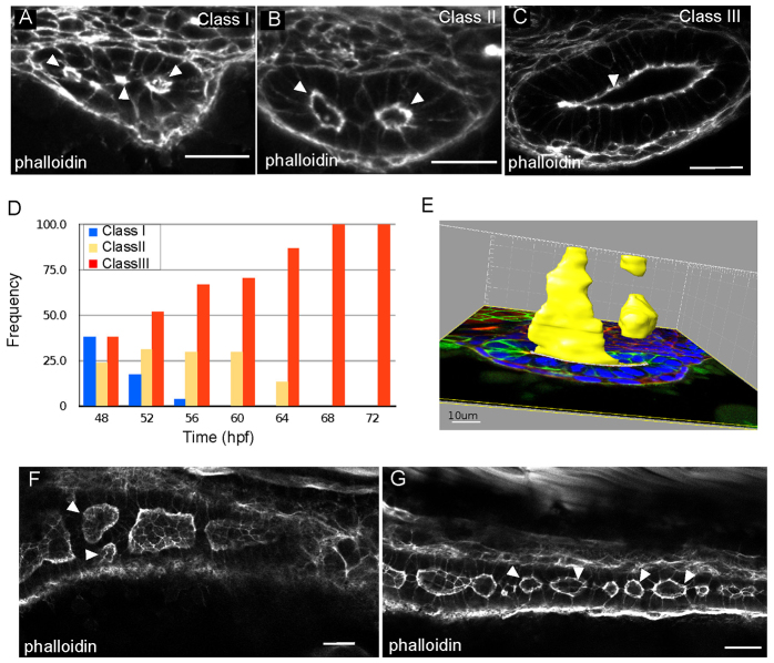
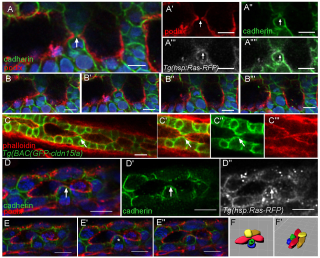
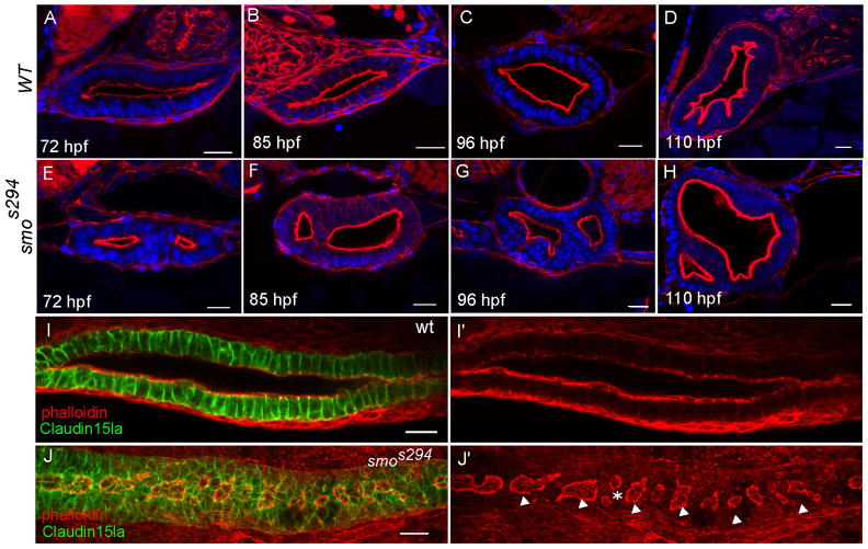
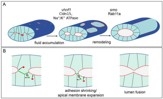
The formation of a single lumen during tubulogenesis is crucial for the development and function of many organs. Although 3D cell culture models have identified molecular mechanisms controlling lumen formation in vitro, their function during vertebrate organogenesis is poorly understood. Using light sheet microscopy and genetic approaches we have investigated single lumen formation in the zebrafish gut. Here we show that during gut development multiple lumens open and enlarge to generate a distinct intermediate, which consists of two adjacent unfused lumens separated by basolateral contacts. We observed that these lumens arise independently from each other along the length of the gut and do not share a continuous apical surface. Resolution of this intermediate into a single, continuous lumen requires the remodeling of contacts between adjacent lumens and subsequent lumen fusion. We show that lumen resolution, but not lumen opening, is impaired in smoothened (smo) mutants, indicating that fluid-driven lumen enlargement and resolution are two distinct processes. Furthermore, we show that smo mutants exhibit perturbations in the Rab11 trafficking pathway and demonstrate that Rab11-mediated trafficking is necessary for single lumen formation. Thus, lumen resolution is a distinct genetically controlled process crucial for single, continuous lumen formation in the zebrafish gut.

Keywords: Lumen; Remodeling; Tubulogenesis.


Figures

Fig. 1.
Fig. 1.
Lumens enlarge and fuse during single lumen formation in the zebrafish gut. (A-C) Confocal images of cross sections of wild-type embryos exhibiting class I (A), class II (B) and class III (C) lumens, stained with phalloidin. Arrowheads indicate the lumens. (D) Quantification of lumen phenotypes between 48 and 72 hpf: 48 hpf n=21, 52 hpf n=29, 56 hpf n=27, 60 hpf n=27, 64 hpf n=30, 68 hpf n=21, 72 hpf n=26. (E) Space-fill projection from a 200 μm confocal stack of an intestine section at the resolution stage. Yellow, lumen; green, GFP-CaaX; blue, DAPI. (F) Confocal whole-mount image of the anterior gut at 58 hpf stained with phalloidin (red). Arrowheads point to adjacent unfused lumens. (G) Confocal whole-mount image of the posterior gut at 58 hpf stained with phalloidin (red). Arrowheads point to lumens. Scale bars: 20 μm in A-C; 10 μm in E; 20 μm in F,G.
Fig. 2.
Fig. 2.
Generation of an intestine-specific transgenic line. (A,B) Dorsal, top panel, and lateral view, bottom panel, in situ hybridization showing claudin 15-like a expressed specifically in the intestine at 56 hpf. (C) Schematic of TgBAC(cldn15la-GFP) generation. The recombination target is shown on top, and the expected protein structure is shown on the bottom. (D) Confocal cross-section of a 72 hpf TgBAC(cldn15la-GFP) embryo. (E) Magnification of box from D. (F) Immunolocalization of Cldn15la to the basolateral membranes of intestinal epithelial cells. (G,H) Whole-mount fluorescent images of 55 hpf and 75 hpf embryos expressing TgBAC(cldn15la-GFP). Arrows indicate the gut tube. (I-L) Live imaging of TgBAC(cldn15la-GFP) using SPIM. Snapshots from a single plane from 48-70 hpf. Arrowheads indicate lumens; arrows point to cell-cell contacts between lumens. (M) Stitched confocal whole-mount images of a TgBAC(cldn15la-GFP) embryo show unfused lumens (arrowhead) in the posterior intestine at 68 hpf that are separated by cell-cell contacts (arrow). (N) Magnification of cell-cell contacts from M. Arrows indicate contacts. Phalloidin (red). Scale bars: 50 μm in D-F; 20 μm in M,N.
Fig. 3.
Fig. 3.
Cell-cell contacts are found between lumens. (A-E) Confocal cross sections of embryos at the lumen resolution stage. (A) Tg(hsp70l:GFP-CaaX) labels all cell membranes. (A′) Cartoon diagram of Fig. 3A depicting laterally arranged lumens in red and ‘bridge’ contacts in green. (B,B′) Apical protein, GFP-Podocalyxin surrounds the lumen but is not found at bridge contacts. Phalloidin (red). (C,C′) Antibody staining against Zo-1 labels tight junctions. Phalloidin (red). (D-E′) Antibody staining against cadherin and β-catenin labels basolateral contacts and ‘bridge’ contacts between lumens. Phalloidin (green). (F) Cartoon depicting two scenarios of lumen fusion along the AP axis. Apical membrane (red) can be deposited on membranes between lumens (top) or lumens may arise isolated and fuse directly without an apical membrane linker (bottom). (G-G′) Whole-mount confocal image of a lumen resolution stage embryo expressing GFP-Podocalyxin (red) and stained for cadherin in green. Cadherin localizes to basolateral contacts separating lumens. Arrows indicate lumens; arrowheads indicate bridge contact. Scale bars: 20 μm.
Fig. 4.
Fig. 4.
Basolateral adhesions separate lumens along the AP axis. (A-A′′) Whole-mount confocal image of an embryo expressing GFP-Podocalyxin (false colored in red) and stained for cadherin (green) shows luminal expansion during a fusion event. Ras-RFP (white) marks cell membranes. Arrows mark fusion event. Blue, DAPI. (B-B′′) Optical sections from a z-stack surrounding a fusion event. (C-C′) Whole-mount confocal image of a TgBAC(GFP-cldn15la) embryo shows a putative adhesion snapping event during fusion. The arrow marks adhesion at the surface. (D-D′) Whole-mount confocal image of an embryo expressing GFP-Podocalyxin (red) and stained for cadherin (green) shows adhesion snapping during fusion. Ras-RFP (white) marks cell membranes. The arrow marks adhesion at the surface. (E-E′) Optical sections from z-stack surrounding a fusion event. The asterisk marks a cell with adhesion. (F,F′) Space-fill projection labeling cells surrounding the fusion event. Lumen, red. The asterisk marks a cell with adhesion. Scale bars: 10 μm.
Fig. 5.
Fig. 5.
smos294 mutants exhibit a lumen fusion defect. (A-H) Confocal cross sections of wild-type (top) and smos294 (bottom) intestines at 72 hpf, 85 hpf, 96 hpf and 110 hpf. Phalloidin (red). (I,J) Confocal whole-mount image of wild-type and smos294 embryos expressing TgBAC(cldn15la-GFP) to highlight the cellular and luminal architecture of the intestine at 72 hpf. (I) Wild-type intestine, (J) smos294 intestine. Arrowheads, lumens; asterisk, adjacent lumens. Scale bars: 20 μm.
Fig. 6.
Fig. 6.
Rab11 is abnormally localized in smos294 mutants. (A,A′) Confocal cross section of a Tg(hsp70l:gal4); Tg(UAS:rab11aS25N) embryo. Phalloidin (green). (B,B′) Confocal cross section of a Tg(hsp70l:rab11bS25N) embryo. Phalloidin (red) (C,D) Confocal cross sections of smos294 and wild-type clutchmates expressing Tg(hsp70l:GFP-RAB11a). Arrowheads point to abnormal enlarged Rab11 positive vesicles in smos294. Phalloidin (red) (E-H) Confocal cross section of wild-type and smos294 embryos expressing Tg(hsp70l:GFP-RAB11a) stained for the apical marker 4e8 (E,F) or cadherin (G,H). Arrowheads point to areas of colocalization. (I,J) Confocal cross sections of uninjected and RFP-Rab11fipa1-injected Tg(hsp70l:GFP-RAB11a) embryos. Arrowheads point to dispersed compartments. (K) Expression levels of Rab11 family members in the intestinal epithelium of smos294 mutants relative to wild-type clutchmates. Rab11fip1a P<0.011, n=3. All embryos are 72 hpf. Scale bars: 20 μm.
Fig. 7.
Fig. 7.
Lumens enlarge and fuse during single lumen formation. (A) During single lumen formation, vhnf1 drives lumen enlargement through Cldn15 and Na+/K+ ATPase regulated fluid accumulation. Next, smoothened regulates remodeling through Rab11a-mediated trafficking to facilitate lumen fusion. Red indicates the luminal surface. (B) During the fusion process, Rab11 traffics apical proteins (red) to the luminal surface and recycles basolateral proteins (green) from bridge contacts to lateral membranes. As the lumens expand, the bridge contacts between the lumens shrink and split, and the lumens fuse.

Similar articles

Cited by

References

    1. Aanstad P., Santos N., Corbit K. C., Scherz P. J., Trinh A., Salvenmoser W., Huisken J., Reiter J. F., Stainier D. Y. R. (2009). The extracellular domain of Smoothened regulates ciliary localization and is required for high-level Hh signaling. Curr. Biol. 19, 1034–1039 - PMC - PubMed
    1. Bagnat M., Cheung I. D., Mostov K. E., Stainier D. Y. R. (2007). Genetic control of single lumen formation in the zebrafish gut. Nat. Cell Biol. 9, 954–960 - PubMed
    1. Bryant D. M., Datta A., Rodríguez-Fraticelli A. E., Peränen J., Martín-Belmonte F., Mostov K. E. (2010). A molecular network for de novo generation of the apical surface and lumen. Nat. Cell Biol. 12, 1035–1045 - PMC - PubMed
    1. Bucci C., Thomsen P., Nicoziani P., McCarthy J., van Deurs B. (2000). Rab7: a key to lysosome biogenesis. Mol. Biol. Cell 11, 467–480 - PMC - PubMed
    1. Chen W., Burgess S., Hopkins N. (2001). Analysis of the zebrafish smoothened mutant reveals conserved and divergent functions of hedgehog activity. Development 128, 2385–2396 - PubMed

Publication types

MeSH terms