Review
. 2014 Feb;20(2):130-8.
doi: 10.1038/nm.3457.

Cell-to-cell transmission of pathogenic proteins in neurodegenerative diseases

Affiliations
Review

Cell-to-cell transmission of pathogenic proteins in neurodegenerative diseases

Jing L Guo et al. Nat Med. 2014 Feb.

Abstract

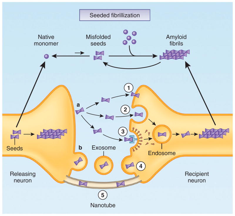
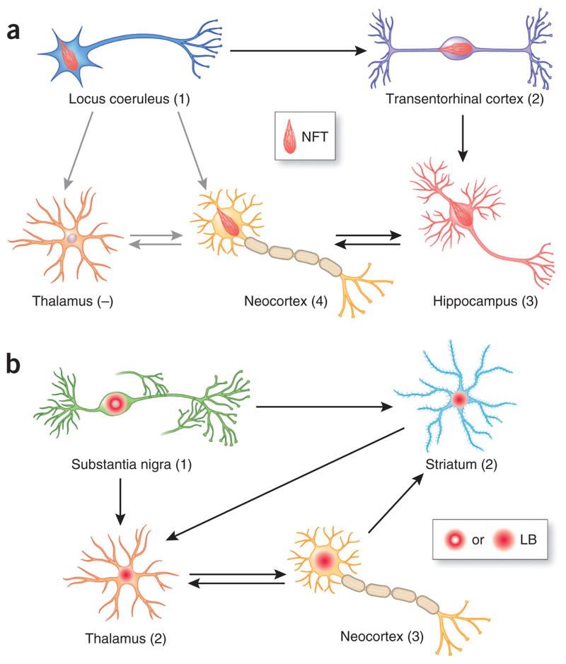
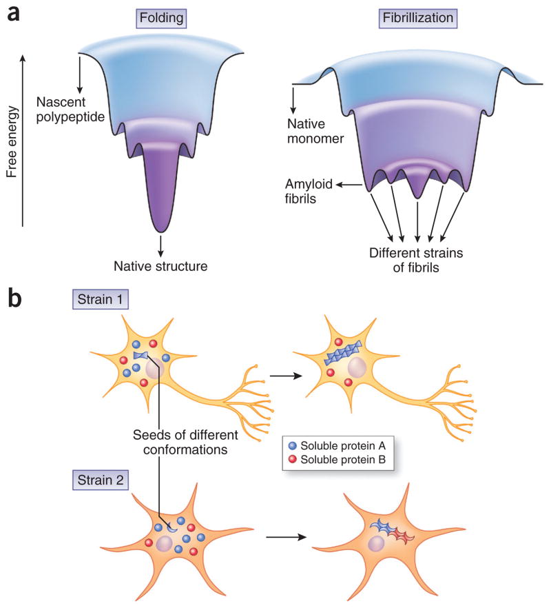
A common feature of many neurodegenerative diseases is the deposition of β-sheet-rich amyloid aggregates formed by proteins specific to these diseases. These protein aggregates are thought to cause neuronal dysfunction, directly or indirectly. Recent studies have strongly implicated cell-to-cell transmission of misfolded proteins as a common mechanism for the onset and progression of various neurodegenerative disorders. Emerging evidence also suggests the presence of conformationally diverse 'strains' of each type of disease protein, which may be another shared feature of amyloid aggregates, accounting for the tremendous heterogeneity within each type of neurodegenerative disease. Although there are many more questions to be answered, these studies have opened up new avenues for therapeutic interventions in neurodegenerative disorders.


Conflict of interest statement

COMPETING FINANCIAL INTERESTS

The authors declare no competing financial interests.

Figures

Figure 1
Figure 1
Potential mechanisms mediating cell-to-cell transmission of cytosolic protein aggregates. (a, b) Misfolded protein seeds (for example, oligomers and protofibrils) first form in the cytoplasm of the releasing neuron (left), where soluble native monomers are recruited into large intracellular aggregates and a positive feedback loop can be initiated by generation of more seeds through fragmentation or secondary nucleation. A small amount of protein aggregates can be released into the extracellular space in the ‘naked’ form (a) or via membrane-bound vesicles such as exosomes (b). Free-floating seeds may directly penetrate the plasma membrane of the recipient neuron (1) or enter by fluid-phase endocytosis (2) or receptor-mediated endocytosis (3), whereas exosomes containing seeds may fuse with the membrane of the recipient neuron (4). Intercellular transfer of seeds may also occur by nanotubes that directly connect the cytoplasm of two cells (5). Internalized seeds then nucleate the fibrillization of native monomers in the cytoplasm of the recipient neuron.
Figure 2
Figure 2
Hypothetical model accounting for the stereotypical progression of pathologies in Alzheimer’s and Parkinson’s diseases. (a, b) Key brain regions developing NFTs (a) or Lewy bodies (LB) (b) are shown for each disease; numbers in parentheses indicate the relative temporal sequence of pathology progression (−, lack of pathology). Major neuronal projections are indicated by arrows, with black arrows indicating projections that hypothetically contribute to the spreading of pathology and grey arrows showing lack of transmission. Brain areas affected by tau and α-syn inclusions at the early stages of the diseases are different, probably owing to brain region–specific vulnerability to the aggregation of the disease proteins. Subsequently affected brain regions may acquire transmissible protein aggregates along both anterograde and retrograde connections, although not all regions connected with affected areas develop pathology. For example, the thalamus is relatively resistant to the accumulation of NFTs despite direct connections with the locus coeruleus. Therefore, brain region–specific vulnerability combined with network connections gives rise to the characteristic onset and progression patterns of neuropathology for different disease proteins.
Figure 3
Figure 3
Pathological strains underlying the divergence and convergence of neurodegenerative proteinopathies. (a) A possible explanation for the existence of multiple fibrillar conformers for a single polypeptide is that the energy landscape of fibrillization is different from that of normal protein folding. Whereas the energy landscape of protein folding resembles a funnel with a global minimum that corresponds to the native structure,, the landscape for abnormal protein aggregation may resemble rugged valleys with numerous local minima corresponding to different strains of fibrils. According to this speculative protein-aggregation landscape, multiple strains of fibrillar aggregates could theoretically emerge in one fibrillization event, but the exact environmental conditions may favor the generation and propagation of one strain over the others; one conformational variant could also directly morph into another if the energy barrier between them is overcome. Indeed, this is what was observed for the Darwinian evolution of prion strains in cultured cells and living animals,. A similar evolution of strains was recently reported for α-syn fibrils generated from recombinant protein, whereby serial passage in vitro resulted in the conversion of one strain of α-syn fibrils into the other. (b) Different strains of amyloid fibrils may exhibit distinct cell tropism (indicated by different shapes of affected cells) and differential efficiency in seeding homotypic monomers (blue spheres) as well as in cross-seeding heterotypic monomers (red spheres). These properties could result in distinct neuropathological profiles, including differences in the distribution of pathology and extent of comorbid pathologies, and ultimately lead to heterogeneous clinical manifestations.

References

    1. Glenner GG, Wong CW. Alzheimer’s disease: initial report of the purification and characterization of a novel cerebrovascular amyloid protein. Biochem Biophys Res Commun. 1984;120:885–890. - PubMed
    1. Kosik KS, Joachim CL, Selkoe DJ. Microtubule-associated protein tau (tau) is a major antigenic component of paired helical filaments in Alzheimer disease. Proc Natl Acad Sci USA. 1986;83:4044–4048. - PMC - PubMed
    1. Spillantini MG, Crowther RA, Jakes R, Hasegawa M, Goedert M. α-synuclein in filamentous inclusions of Lewy bodies from Parkinson’s disease and dementia with Lewy bodies. Proc Natl Acad Sci USA. 1998;95:6469–6473. - PMC - PubMed
    1. Neumann M, et al. Ubiquitinated TDP-43 in frontotemporal lobar degeneration and amyotrophic lateral sclerosis. Science. 2006;314:130–133. - PubMed
    1. DiFiglia M, et al. Aggregation of hunting tin in neuronal intranuclear inclusions and dystrophic neurites in brain. Science. 1997;277:1990–1993. - PubMed

Publication types

MeSH terms

Substances