Review
. 2014 Jun;39(3):588-604.
doi: 10.1007/s00261-014-0083-3.

Renal angiomyolipoma: a radiological classification and update on recent developments in diagnosis and management

Affiliations
Review

Renal angiomyolipoma: a radiological classification and update on recent developments in diagnosis and management

Masahiro Jinzaki et al. Abdom Imaging. 2014 Jun.

Abstract

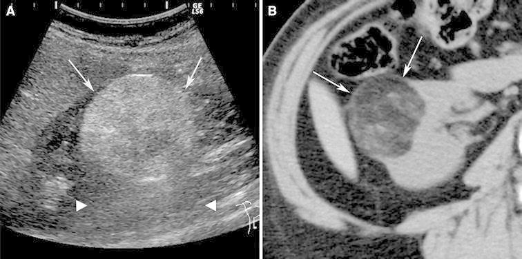
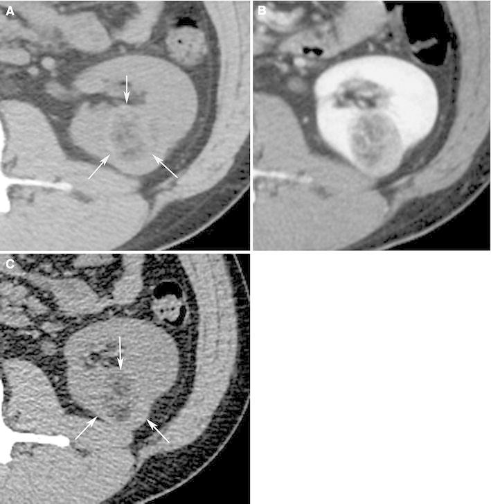
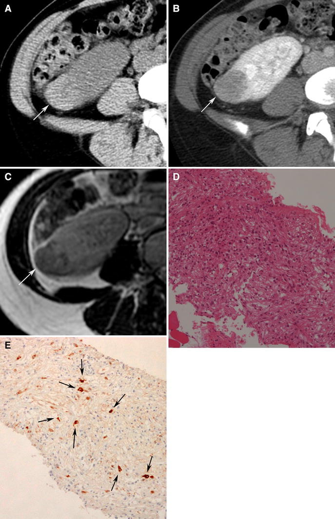
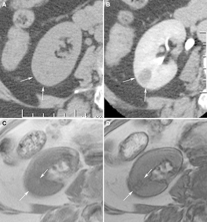
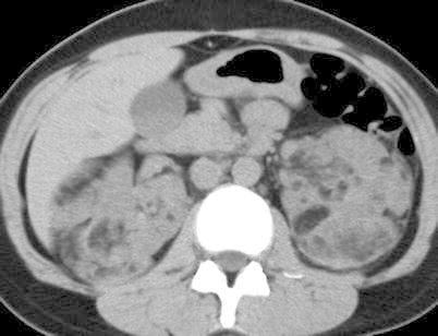
Angiomyolipoma is the most common benign solid renal neoplasm observed in clinical practice. Once thought to be a hamartoma and almost always diagnosed by the imaged-based detection of fat, angiomyolipomas are now known to consist of a heterogeneous group of neoplasms. Although all are considered perivascular epithelioid cell tumors, many display different pathology, imaging features, and clinical behavior. The importance of understanding this group of neoplasms is emphasized by the fact that many types of angiomyolipoma contain little to no fat, and despite being benign, sometimes escape a pre-operative diagnosis. These types of angiomyolipomas can all be considered when encountering a renal mass that is both hyperattenuating relative to renal parenchyma on unenhanced CT and T2-hypointense, features that reflect their predominant smooth muscle component. We review recent developments and provide a radiological classification of angiomyolipomas that helps physicians understand the various types and learn how to both diagnose and manage them.


Figures

Fig. 1
Fig. 1
Angiomyolipoma, classic type, in a 61-year-old woman. Ultrasound (A) shows a 4.2-cm right renal mass (arrows) that is markedly hyperechoic relative to renal parenchyma and accompanied by acoustic shadowing (arrow heads). Transverse, unenhanced CT image (5-mm sections) shows a right renal mass (arrows) with fat attenuation (−60 HU).
Fig. 2
Fig. 2
Angiomyolipoma, classic type, with small amount of fat in a 40-year-old man. Transverse, unenhanced CT (A) and enhanced (B) images (both using 5-mm sections) shows a 3.4-cm left renal mass (arrows) attenuation values that were measured on unenhanced images were all higher than −10 HU. When 1.5-mm sections were reconstructed (C), an attenuation of −25 HU was obtained and the diagnosis of angiomyolipoma was made. Fat attenuation was identified only on unenhanced CT.
Fig. 3
Fig. 3
Angiomyolipoma, fat poor type, hyperattenuating subtype in a 39-year-old woman. Transverse, unenhanced CT (5-mm sections) demonstrates a 2.9-cm hyperattenuating (47 HU) right renal mass (arrows) with no regions that measured less than 30 HU (A). The mass-enhanced homogenously at CT (B) and was hypointense on transverse T2-weighted image (fast spin-echo, TR:2400, TE:94) (C). Biopsy specimen showed smooth muscle component (hematoxylin–eosin stain; original magnification, ×100.) (D), and was positive for HMB-45 (E: arrows).
Fig. 4
Fig. 4
Angiomyolipoma, fat poor, isoattenuating subtype in a 71-year-old woman. Transverse, unenhanced CT (5 mm sections) demonstrates a 1.8-cm isoattenuating right renal mass (arrows) with an attenuation of 33HU; no regions measured less than -6 HU (A). The mass enhanced to an attenuation of 78HU (B). MRI signal intensity of the tumor was 70 on in-phase image (fast spoiled gradient echo, TR:125, TE:4.2) (C: arrows), and 39 on opposed phase image (fast spoiled gradient echo, TR:160, TE:2.0) (D: arrows). The mass was hypointense on T2-weighted imaging (single shot fast spin echo, TR:26000, TE:189) (E). Because this enhancing renal mass was isoattenuating, T2-hypointense, and suppressed on chemical shift MR imaging, isoattenuating angiomyolipoma was suspected and proven by percutaneous biopsy. Hemotoxlyin-eosin stain specimen (F) showed smooth muscle component (arrow) and scattered fat cells (bold arrows).
Fig. 4
Fig. 4
Angiomyolipoma, fat poor, isoattenuating subtype in a 71-year-old woman. Transverse, unenhanced CT (5 mm sections) demonstrates a 1.8-cm isoattenuating right renal mass (arrows) with an attenuation of 33HU; no regions measured less than -6 HU (A). The mass enhanced to an attenuation of 78HU (B). MRI signal intensity of the tumor was 70 on in-phase image (fast spoiled gradient echo, TR:125, TE:4.2) (C: arrows), and 39 on opposed phase image (fast spoiled gradient echo, TR:160, TE:2.0) (D: arrows). The mass was hypointense on T2-weighted imaging (single shot fast spin echo, TR:26000, TE:189) (E). Because this enhancing renal mass was isoattenuating, T2-hypointense, and suppressed on chemical shift MR imaging, isoattenuating angiomyolipoma was suspected and proven by percutaneous biopsy. Hemotoxlyin-eosin stain specimen (F) showed smooth muscle component (arrow) and scattered fat cells (bold arrows).
Fig. 5
Fig. 5
Angiomyolipoma with epithelial cysts in a 60-year-old man. Transverse, unenhanced CT image (1.5 mm sections) shows a 2.0-cm left renal mass with central, hypoaattenuating (5 HU) and a peripheral hyperattenuating (50 HU) components (A: arrow). The peripheral component enhanced (B: arrow). This mass was resected during nephrectomy for an ipsilateral renal cell carcinoma. Pathology revealed angiomyolipoma with an epithelial-lined cyst (C: arrowheads). The cystic area on pathologic specimencorrespond to the low attenuation area seen on CT.
Fig. 6
Fig. 6
Angiomyolipoma with epithelial cysts in a 46-year-old man. Transverse, T2-weighted MR image (fast spin-echo, TR:4000, TE:92) shows a 2.5-cm multilocular cystic mass with a hyperintense central component, and hypointense wall (A, B: arrows) and septa (B: arrowhead). Because of suspicion for renal cell carcinoma, this mass was resected. The specimen showed angiomyolipoma with epithelial cysts; the wall contained smooth muscle component.
Fig. 7
Fig. 7
Epithelioid angiomyolipoma in a 21-year-old man. Transverse, unenhanced CT (5 mm sections) shows a 12.0 cm hyperattenuating right renal mass (A: arrows) that enhanced heterogeneously (B: arrows). No regions of fat attenuation could be identified. Hematoxylin–eosin staining specimens show pleomorphic tumor cells with large hyperchromatic nuclei and abundant eosinophilic cytoplasm (C). The tumor cells were was positive for HMB-45 (D arrows).
Fig. 8
Fig. 8
Epithelioid angiomyolipomae in a 40-year-old woman. Transverse, unenhanced CT (5-mm sections) (A) and enhanced CT (B) demonstrates a 5.0-cm multilocular cystic mass in the left kidney. Both the wall and septa (arrows) were hyperattenuating (48 HU) and enhanced. On transverse, T2-weighted MR image (fat-saturated fast spin-echo, TR:3800, TE: 89) (C), the wall (arrows) and septum (arrowhead) of the multilocular cystic mass appeared hypointense. On a transverse, T1-weighted MR image (fast spoiled gradient echo, TR: 150, TE:2.3) (D), the cystic component was hyperintense (arrows). The mass was considered a Bosniak Category 3 lesion and resected due to its appearance. The hyperintense cystic component corresponded to hemorrhage at pathology.
Fig. 9
Fig. 9
Angiomyolipoma in a 32-year-old woman with tuberous sclerosis complex. Transverse, unenhanced CT (5-mm sections) shows multiple bilateral renal masses each containing fat attenuation (less than −10 HU) diagnostic of angiomyolipomas.

References

    1. Eble JN, Sauter G, Epstein JI, Sesterhenn IA. World Health Organization classification of tumors: pathology and genetics. Tumors of the urinary system and male genital organs. Lyon: IARC Press; 2004.
    1. Lane BR, Aydin H, Danforth TL, et al. Clinical correlates of renal angiomyolipoma subtypes in 209 patients: classic, fat poor, tuberous sclerosis associated and epithelioid. J Uro. 2008;180:836–843. doi: 10.1016/j.juro.2008.05.041. - DOI - PubMed
    1. Tamboli P, Ro JY, Amin MB, Ligato S, Ayala AG. Benign tumors and tumor-like lesions of the adult kidney. Part II: benign mesenchymal and mixed neoplasms, and tumor-like lesions. Adv Anat Pathol. 2000;7:47–66. doi: 10.1097/00125480-200007010-00007. - DOI - PubMed
    1. Milner J, McNeil B, Alioto J, et al. Fat poor renal angiomyolipoma: patient, computerized tomography and histological findings. J Urol. 2006;176:905–909. doi: 10.1016/j.juro.2006.04.016. - DOI - PubMed
    1. Jinzaki M, Tanimoto A, Narimatsu Y, et al. Angiomyolipoma: imaging findings in lesions with minimal fat. Radiology. 1997;205:497–502. - PubMed

MeSH terms

LinkOut - more resources