

Expanding mouse ventricular cardiomyocytes through GSK-3 inhibition
- PMID: 24505027
- PMCID: PMC3934298
- DOI: 10.1002/0471143030.cb2309s61
Expanding mouse ventricular cardiomyocytes through GSK-3 inhibition
Abstract
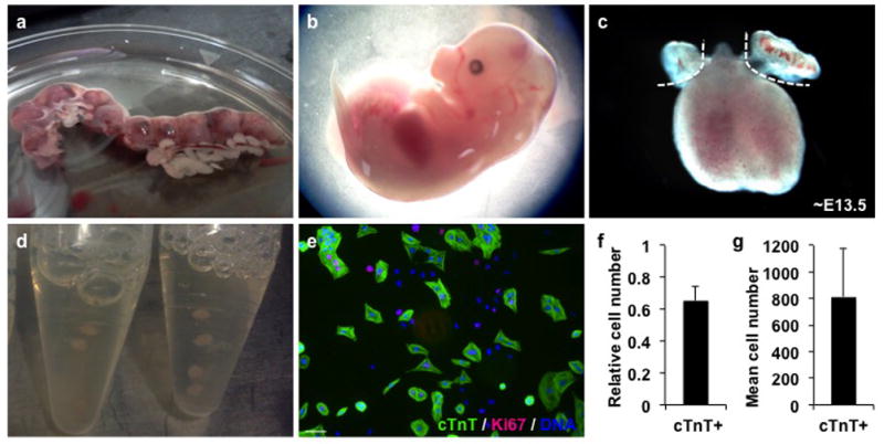
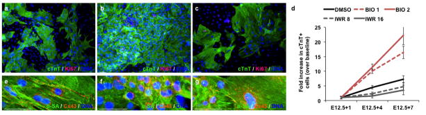
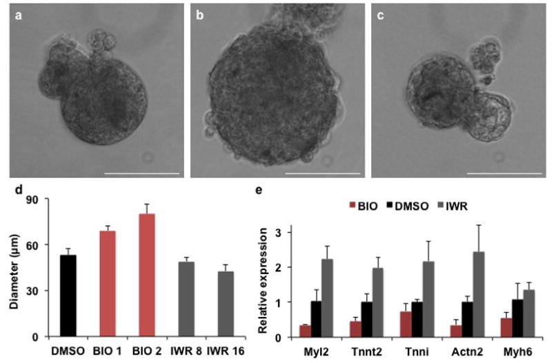
Controlled proliferation of cardiomyocytes remains a major limitation in cell biology and one of the main underlying hurdles for true modern regenerative medicine. Here, a technique is described for robust expansion of early fetal-derived mouse ventricular cardiomyocytes on a platform usable for high-throughput molecular screening, tissue engineering and, potentially, in vivo translational experiments. This method provides a small-molecule approach to control proliferation or differentiation of early beating cardiomyocytes through modulation of the Wnt/β-catenin signaling pathway. Moreover, isolation and expansion of fetal cardiomyocytes takes less than 3 weeks, yields a relatively pure (∼70%) functional myogenic population, and is highly reproducible.
Keywords: GSK-3 inhibitor; Wnt/β-catenin signaling; cardiomyocyte proliferation; differentiation; expansion; isolation.
Copyright © 2013 John Wiley & Sons, Inc.
Figures
References
-
- Akins RE, Boyce RA, Madonna ML, Schroedl NA, Gonda SR, McLaughlin TA, Hartzell CR. Cardiac organogenesis in vitro: reestablishment of three-dimensional tissue architecture by dissociated neonatal rat ventricular cells. Tissue Eng. 1999;5:103–118. - PubMed
-
- Buckingham M, Meilhac S, Zaffran S. Building the mammalian heart from two sources of myocardial cells. Nat Rev Genet. 2005;6:826–835. - PubMed
-
- Bursac N, Papadaki M, White JA, Eisenberg SR, Vunjak-Novakovic G, Freed LE. Cultivation in rotating bioreactors promotes maintenance of cardiac myocyte electrophysiology and molecular properties. Tissue Eng. 2003;9:1243–1253. - PubMed
Publication types
MeSH terms
Substances
Grants and funding
LinkOut - more resources
Full Text Sources
Other Literature Sources
