

Macular hole formation after pars plana vitrectomy for the treatment of Valsalva retinopathy: a case report
- PMID: 24505205
- PMCID: PMC3913987
- DOI: 10.3341/kjo.2014.28.1.91
Macular hole formation after pars plana vitrectomy for the treatment of Valsalva retinopathy: a case report
Abstract
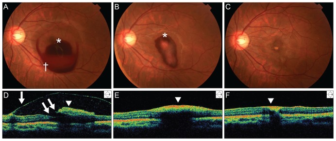
We report a case of complete surgical resolution of Valsalva retinopathy that manifested as a premacular hemorrhage involving a membrane followed by a macular hole (MH) resulting from the first vitrectomy. A 20-year-old female patient was referred to our hospital due to sudden vision loss in the left eye. Her best-corrected visual acuity (BCVA) in the left eye was hand motion. Fundus photographs and optical coherence tomography (OCT) revealed a premacular hemorrhage. Nine weeks later, the BCVA in the left eye had returned to 20 / 100 and the premacular hemorrhage had completely resolved, but residual sub-internal limiting membrane deposits and a preretinal membrane were present. The preretinal membrane was removed by core vitrectomy and preretinal membrane peeling, but the foveal deposits could not be excised. Two weeks after the first vitrectomy, the deposits resolved spontaneously, but a full-thickness MH was present. Six months after a second vitrectomy with fluid-gas exchange, the BCVA in the left eye had improved to 20 / 25 and OCT showed that the MH had closed. This case illustrates the possibility of MH formation following vitrectomy for Valsalva retinopathy.
Keywords: Macular hole; Valsalva retinopathy; Vitrectomy.
Conflict of interest statement
No potential conflict of interest relevant to this article was reported.
Figures


References
-
- Duane TD. Valsalva hemorrhagic retinopathy. Am J Ophthalmol. 1973;75:637–642. - PubMed
-
- Gass JD. Stereoscopic atlas of macular diseases: diagnosis and treatment. 4th ed. St. Louis: Mosby; 1997. pp. 752–754.
-
- Roberts DK, MacKay KA. Microhemorrhagic maculopathy associated with aerobic exercise. J Am Optom Assoc. 1987;58:415–418. - PubMed
-
- Friberg TR, Braunstein RA, Bressler NM. Sudden visual loss associated with sexual activity. Arch Ophthalmol. 1995;113:738–742. - PubMed
-
- De Crecchio G, Pacente L, Alfieri MC, Greco GM. Valsalva retinopathy associated with a congenital retinal macrovessel. Arch Ophthalmol. 2000;118:146–147. - PubMed
Publication types
MeSH terms
LinkOut - more resources
Full Text Sources
Other Literature Sources
Medical
Miscellaneous
