Spontaneous rupture of intracranial dermoid tumor in a patient with vertigo. Computed tomography and magnetic resonance imaging findings
- PMID: 24505228
- PMCID: PMC3908513
- DOI: 10.12659/PJR.889172
Spontaneous rupture of intracranial dermoid tumor in a patient with vertigo. Computed tomography and magnetic resonance imaging findings
Abstract
Background: Congenital dermoid cysts are very rare, constituting less than 1% of intracranial tumors. Spontaneous rupture of dermoid tumor is a potentially serious complication that can lead to meningitis, seizures, cerebral ischemia and hydrocephalus. Occasionally, dermoid tumors are incidentally discovered on computed tomography (CT) of the brain or magnetic resonance imaging (MRI) following unrelated clinical complaints. They are also discovered during radiologic investigations of unexplained headaches, seizures, and rarely olfactory delusions.
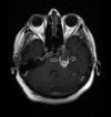
Case report: In this report we describe a patient complaining of vertigo caused by spontaneous rupture of dermoid cyst, preoperatively diagnosed by CT and MRI. Cranial CT revealed a dense fatty lesion adjacent to the posterolateral parasellar region on the left with multiple small, dense fat droplets scattered in the subarachnoid space corresponding to a dermoid cyst rupture. Cranial MRI sections revealed a lesion with mixed-signal-intensity and multiple hyperintense droplets scattered through the cerebellar surface on the left. No enhancement was found on axial T1-weighted MRI after intravenous Gadolinium administration. Diffusion weighted image (DWI) and apparent diffusion coefficient map studies exhibited explicit restricted diffusion.
Discussion: Many studies and literature case reports concerning the rupture of dermoid cyst have been reported. However, multimodal imaging of this rare pathology in the same patient is uncommon. Although dermoid cysts are pathognomonic in appearance on a CT examination, the MRI is also of value in helping to understand the effect of extension and pressure of the mass. DWI is also important for support of the diagnosis and patient follow-up.
Keywords: computed tomograph; dermoid tumor; magnetic resonance imaging; rupture; vertigo.
Figures


References
-
- Cha JG, Paik SH, Park JS, et al. Ruptured spinal dermoid cyst with disseminated intracranial fat droplets. Br J Radiol. 2006;79:167–69. - PubMed
-
- Venkatesh SK, Phadke RV, Trivedi P, et al. Asymptomatic spontaneous rupture of suprasellar dermoid cyst: a case report. Neurol India. 2002;50:480–83. - PubMed
-
- Osborn AG, Preece MT. Intracranial cysts: radiologic-pathologic correlation and imaging approach. Radiology. 2006;239:650–64. - PubMed
Publication types
LinkOut - more resources
Full Text Sources
Other Literature Sources
