. 2014 Jan 29;4(3):316-35.
doi: 10.7150/thno.7819. eCollection 2014.

Potential theranostics application of bio-synthesized silver nanoparticles (4-in-1 system)

Affiliations

Potential theranostics application of bio-synthesized silver nanoparticles (4-in-1 system)

Sudip Mukherjee et al. Theranostics. .

Abstract

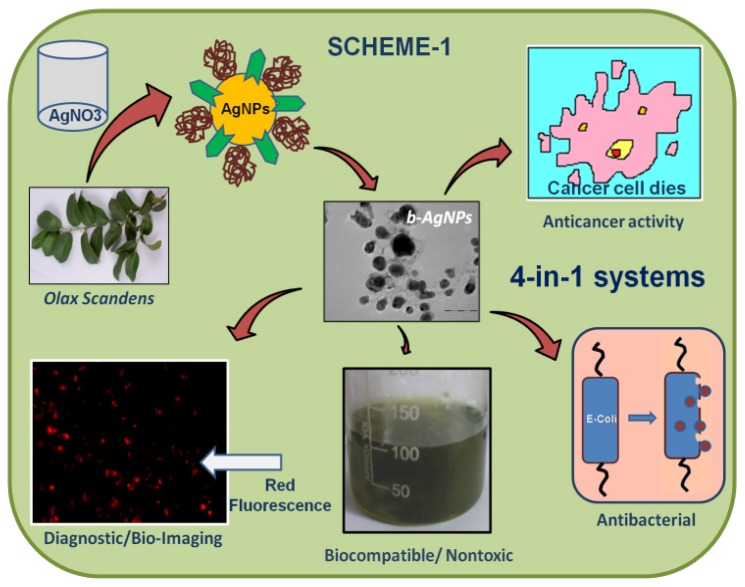
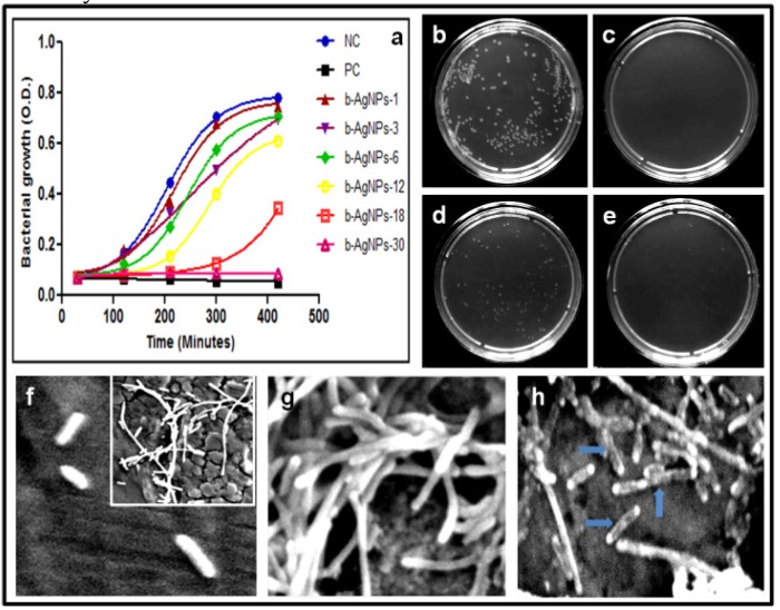
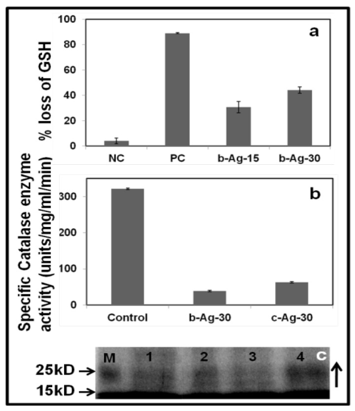
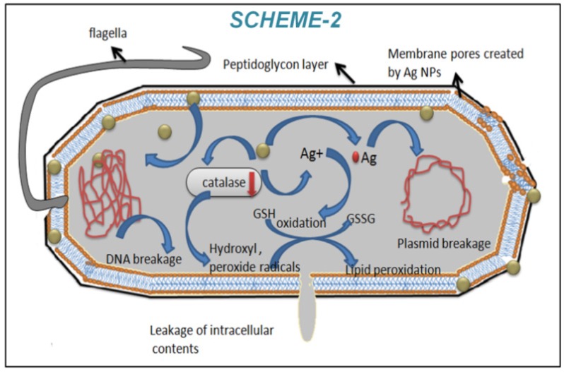
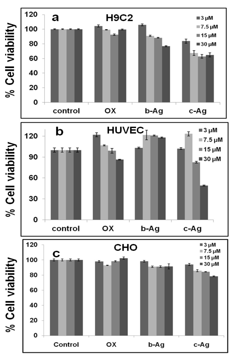
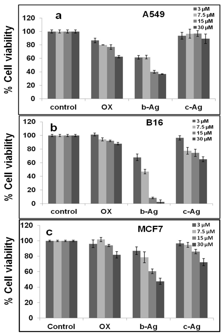
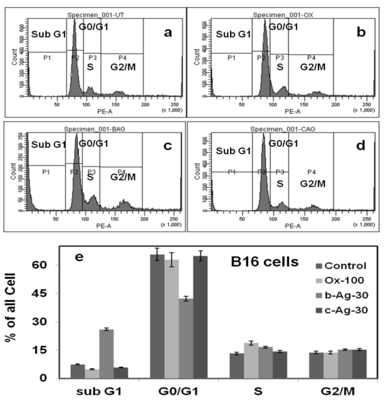
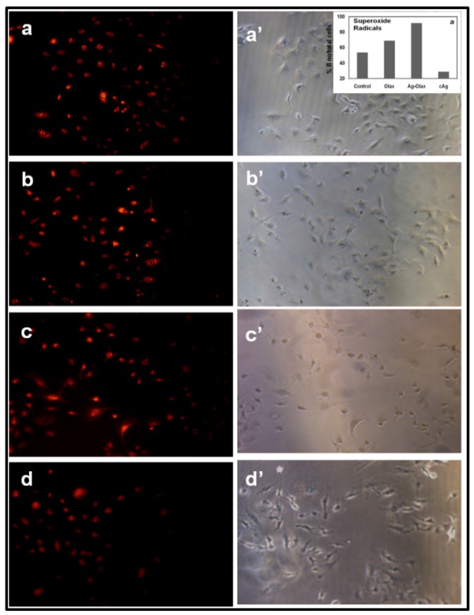
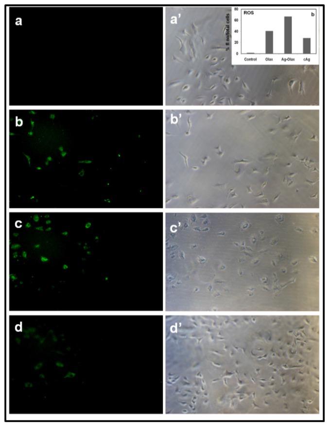
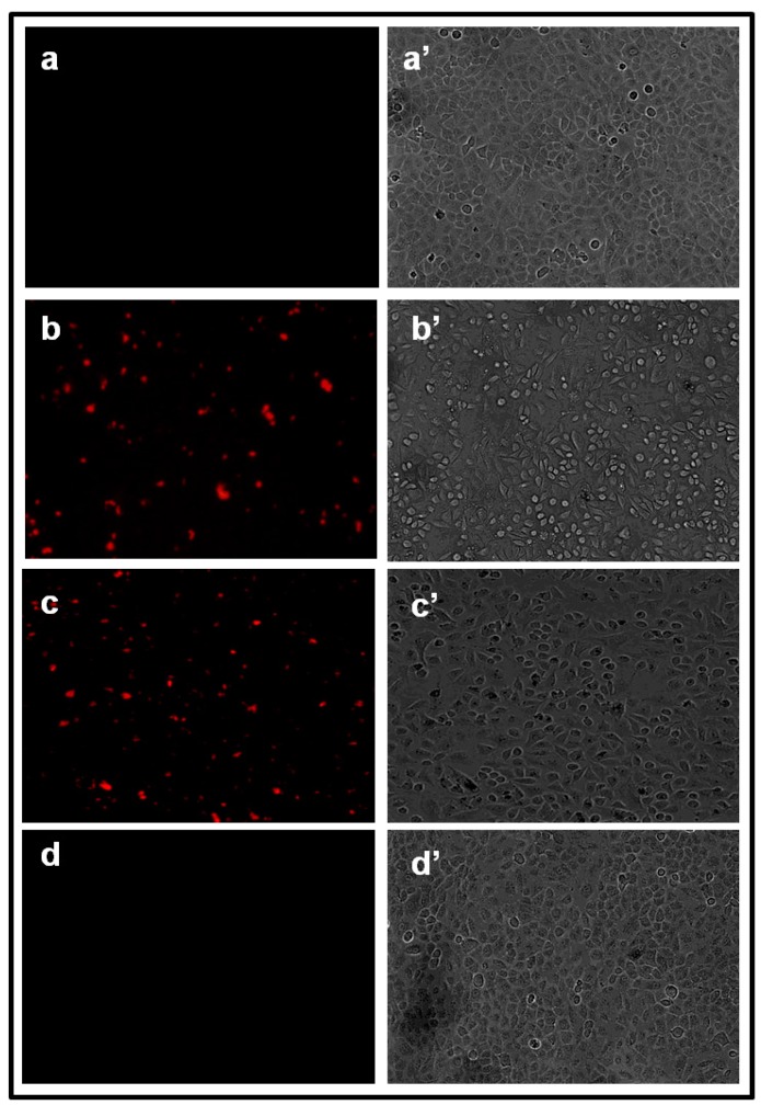
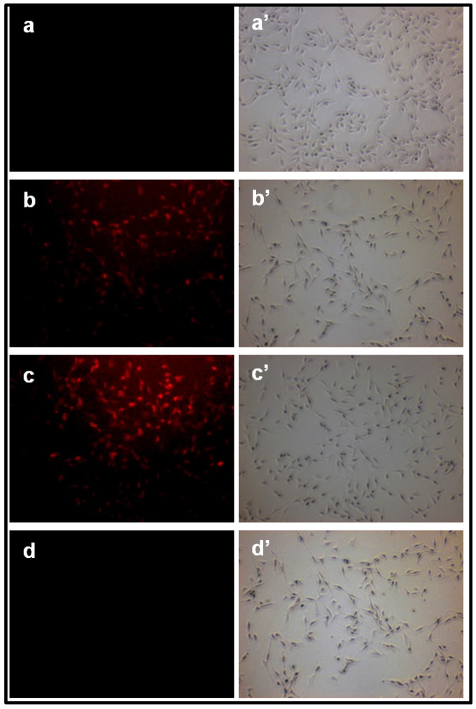
In this report, we have designed a simple and efficient green chemistry approach for the synthesis of colloidal silver nanoparticles (b-AgNPs) that is formed by the reduction of silver nitrate (AgNO3) solution using Olax scandens leaf extract. The colloidal b-AgNPs, characterized by various physico-chemical techniques exhibit multifunctional biological activities (4-in-1 system). Firstly, bio-synthesized silver nanoparticles (b-AgNPs) shows enhanced antibacterial activity compared to chemically synthesize silver nanoparticles (c-AgNPs). Secondly, b-AgNPs show anti-cancer activities to different cancer cells (A549: human lung cancer cell lines, B16: mouse melanoma cell line & MCF7: human breast cancer cells) (anti-cancer). Thirdly, these nanoparticles are biocompatible to rat cardiomyoblast normal cell line (H9C2), human umbilical vein endothelial cells (HUVEC) and Chinese hamster ovary cells (CHO) which indicates the future application of b-AgNPs as drug delivery vehicle. Finally, the bio-synthesized AgNPs show bright red fluorescence inside the cells that could be utilized to detect the localization of drug molecules inside the cancer cells (a diagnostic approach). All results together demonstrate the multifunctional biological activities of bio-synthesized AgNPs (4-in-1 system) that could be applied as (i) anti-bacterial & (ii) anti-cancer agent, (iii) drug delivery vehicle, and (iv) imaging facilitator. To the best of our knowledge, there is not a single report of biosynthesized AgNPs that demonstrates the versatile applications (4-in-1 system) towards various biomedical applications. Additionally, a plausible mechanistic approach has been explored for the synthesis of b-AgNPs and its anti-bacterial as well as anti-cancer activity. We strongly believe that bio-synthesized AgNPs will open a new direction towards various biomedical applications in near future.

Keywords: Antibacterial; Bio-synthesis; Green Chemistry; Multifunctional activities; Olax scandens; Silver nanoparticle; anti-cancer..


Conflict of interest statement

Competing Interests: The authors have declared that no competing interest exists.

Figures

Figure A
Figure A
(Scheme 1) Over all presentation for synthesis, characterization and biomedical applications (diagnostic, anticancer antibacterial applications) of biosynthesized silver nanoparticles (b-AgNPs) using Olax Scandens leaf extract.
Figure 1
Figure 1
Physico-chemical characterization and stability studies of b-AgNPs. (a) absorbance of b-AgNPs obtained after 24 hours with different concentration of Olax extract has been measured by UV visible spectroscopy (150, 400, 500 & 750 values indicate the volume of Olax extract in µL), (b) X-ray diffraction pattern (XRD) of b-AgNPs-500 demonstrates the crystalline nature of silver nanoparticles obtained with 500 µL of extract, (c) TEM indicates the morphology of b-AgNPs-500 and (d) in-vitro stability studies of as synthesized b-AgNPs-500 (24 h to 14 days) has been carried out to several physiological buffer and salts solution at different pH. The results indicate the high stability of b-AgNPs.
Figure 2
Figure 2
Study of anti-bacterial activities: (a) liquid growth inhibition kinetics of E. coli using different concentrations of b-AgNPs. b-AgNP-30 (at 30 µM) shows almost 100% growth inhibition. Ampicillin has been used as a positive control (PC) & NC: negative control or untreated E. coli. The numerical number indicates the concentration of b-AgNPs in µM, (b-e) optical images of bacterial colonies formed by E. coli cells i.e. colony counting assay (after 24 h): b: Control, c:Ampicillin (100μg/ml), d: b-AgNPs (18 μM), e: b-AgNPs (30μM) and (f-h) SEM images of E. coli cells (f) without being treated (control), (g) treated with Olax for 1 hour, (h) treated with b-AgNPs (30 µM) for 1 hour. The SEM images show the silver nanoparticles damages the bacterial cell membrane (marked by blue arrow), whereas, the bacterial membranes of untreated and treated E. coli with Olax is intact.
Figure 3
Figure 3
Study of oxidative stress and SDS PAGE. (a) In-vitro GSH oxidation by Ellman's assay: PC: positive control (1 mM H2O2), and b-AgNPs-15 and b-AgNPs-30: biosynthesized silver nanoparticles synthesized by Olax leaf extract (15 & 30 indicates µM). Results show that b-AgNPs (in both the concentrations) shows considerable amount of oxidation of glutathione, which generates oxidative stress inside bacteria cell. (b) In vitro catalase activity: Control, biosynthesized b-AgNPs (30 µM) treated E. coli and chemically synthesized c-AgNPs (30 µM) treated E. coli. Biosinthesized AgNPs show maximum inhibition effect. (c) Coomassie blue staining of SDS page of bacterial proteins: 1D gel electrophoresis study of E. coli proteins treated with (2) Ampicillin (100μg/ml), (3) c-AgNPs (30 µM) and b-AgNPs (30 µM). M: standard protein marker and (1) untreated E. coli. Bacteria cells treated with b-AgNPs show the up regulation of stress proteins (10 kD-25 kD) in b-AgNPs (30 µM).
Figure B
Figure B
(Scheme 2) Schematic presentation of antibacterial activity of b-AgNPs towards E. coli using biosynthesized silver nanoparticles.
Figure 4
Figure 4
Cell viability assay using MTT reagents. In vitro anti proliferative assay by MTT reagent in (a) H9C2, (b) HUVEC & (c) CHO. The numerical value represents the concentrations in µM for respective samples. Biosynthesized silver nanoparticles, biocompatible towards H9C2, HUVEC & CHO. *All values are significant, i.e. P < 0.05.
Figure 5
Figure 5
Cell viability assay using MTT reagents. In vitro anti proliferative assay by MTT reagent in (a) A549, (d) B16 and (c) MCF7 cell lines for 24 hours in dose dependent manner. Biosynthesized silver nanoparticles (b-AgNPs) and chemically synthesized AgNPs (c-AgNPs) have been given as treatment as different doses. The numerical value represents the concentrations in µM for respective samples. Biosynthesized silver nanoparticles show anti-cancer activity towards A549 and B16 cancer cell lines. *All values are significant, i.e. P < 0.05.
Figure 6
Figure 6
Cell cycle assay using PI-RNase reagents in B16 cells. Extent of propidium iodide staining of the gated population was displayed in a histogram and the regions are defined as: sub-G1, G0/G1, S, and G2/M. Result shows the sub-G1 cell accumulation for b-Ag-30 treated B16 melanoma cancer cells indicates the apoptotic cell arrests or death.
Figure 7
Figure 7
Formation of superoxide anion inside A549 cancer cell measured by fluorescence microscopy: Fluorescence images of A549 cells treated with (a) nothing; negative control, (b) Olax extract (100 µg/ ml), (c) b-AgNPs at 30 µM and (d) c-AgNPs at 30 µM, respectively. Images of a'-d' correspond to bright-filed phase images. Biosynthesized AgNPs support the formation of super oxide anion inside the A549 cells. Insets of figure.5.a'. shows the quantitative analysis of % (total number of fluorescence/ cell numbers).
Figure 8
Figure 8
Formation of hydrogen peroxide inside A549 cancer cell measured by fluorescence microscopy: Fluorescence images of A549 cells treated with (a) nothing; negative control, (b) Olax extract (100 µg/ ml), (c) b-AgNPs at 30 µM and (d) c-AgNPs at 30 µM, respectively. Images of a'-d' correspond to bright-filed phase images. Biosynthesized AgNPs support the formation of hydrogen peroxide inside the A549 cells. Insets of figure.6.a'. shows the quantitative analysis of % (total number of fluorescence/ cell numbers).
Figure 9
Figure 9
a-b: Western blot analysis of b-AgNPs treated B16 cells show upregulation of p53 and active Caspase-3; (b) Quantification of signal intensity of each of the protein was normalized with the corresponding β-Actin signal shows increase in p53 & active caspase-3.
Figure 10
Figure 10
a-b: (a) atomic absorption spectroscopy to quanitify the amount of silver ion released in acidic and physiological pH, (b) UV-Visible studies show that the comparitive UV spectrum of b-AgNPs in acidic and physiological pH solution.
Figure 11
Figure 11
Fluorescence and the corresponding phase images of untreated A549 cells and cells treated with Olax, b-AgNPs c-AgNPs, observed by an Olympus Fluorescence Microscope. Fluoresence images of A549 cells treated with (a) untreated or control, (b) Olax (100 µg/ ml) leaf extract ,(c) b-AgNPs (at 30 µM) and (d) c-AgNPs (at 30 µM). Images of a', b', c' & d' correspond to phase images. All the treated A549 cells were extensively washed with DPBS (6 times) before taking the fluorescence images. It is to be noted that there is no significant cell killing is observed at 30 µM.
Figure 12
Figure 12
Fluorescence and the corresponding phase images of untreated B16 cells and cells treated with Olax, b-AgNPs c-AgNPs, observed by an Olympus Fluorescence Microscope. Fluoresence images of B16 cells treated with (a) untreated or control, (b) Olax (100 µg/ ml) leaf extract ,(c) b-AgNPs (at 30 µM) and (d) c-AgNPs (at 30 µM). Images of a', b', c' & d' correspond to phase images. All the treated B16 cells were extensively washed with DPBS (6 times) before taking the fluorescence images. It is to be noted that there is no significant cell killing is observed at 30 µM.

Similar articles

Cited by

References

    1. Alivisatos P. The use of nanocrystals in biological detection. Nature Biotechnology. 2004;22:47–52. - PubMed
    1. Salata O. Applications of nanoparticles in biology and medicine. J Nanobiotechnology. 2004;2(1):3.. Epub 2004/05/04. doi: 10.1186/1477-3155-2-3. - PMC - PubMed
    1. Gao X, Cui Y, Levenson RM, Chung LWK, Nie S. In vivo cancer targeting and imaging with semiconductor quantum dots. Nature Biotechnology. 2004;22:969–76. - PubMed
    1. Giljohann DA, Seferos DS, Daniel WL, Massich MD, Patel PC, Mirkin CA. Gold Nanoparticles for Biology and Medicine. Angew Chem Int Ed. 2010;49:3280–94. - PMC - PubMed
    1. Patra CR, Bhattacharya R, Patra S, Vlahakis NE, Gabashvili A, Koltypin Y. et al. Pro-angiogenic properties of europium(III) hydroxide nanorods. Advanced Materials. 2008;20(4):753–6.

Publication types